Document 14111226
advertisement
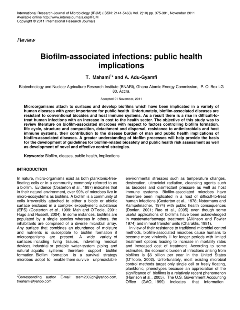
International Research Journal of Microbiology (IRJM) (ISSN: 2141-5463) Vol. 2(10) pp. 375-381, November 2011 Available online http://www.interesjournals.org/IRJM Copyright © 2011 International Research Journals Review Biofilm-associated infections: public health implications T. Mahami1* and A. Adu-Gyamfi Biotechnology and Nuclear Agriculture Research Institute (BNARI), Ghana Atomic Energy Commission, P. O. Box LG 80, Accra. Accepted 01 November, 2011 Microorganisms attach to surfaces and develop biofilms which have been implicated in a variety of human diseases with great importance for public health .Unfortunately, biofilm-associated diseases are resistant to conventional biocides and host immune systems. As a result there is a rise in difficult-totreat human infections with an increase in cost to the health sector. The objective of this study was to review literature on biofilm-associated microbes with respect to factors controlling biofilm formation, life cycle, structure and composition, detachment and dispersal, resistance to antimicrobials and host immune systems, their contribution to the disease burden of man and public health implications of biofilm-associated diseases. A greater understanding of biofilm processes will help provide the basis for the development of guidelines for biofilm-related biosafety and public health risk assessment as well as development of novel and effective control strategies. Keywords: Biofilm, dieases, public health, implications INTRODUCTION In nature, micro-organisms exist as both planktonic-freefloating cells or in a community commonly referred to as a biofilm. Evidence (Costerton et al., 1987) indicates that in their natural environment, over 99% of microbes live in micro-ecosystems as biofilms. A biofilm is a community of cells irreversibly attached to either a biotic or abiotic surface enclosed in a complex exopolymeric substance (EPS) (Costerton et al., 1999: Mah and O’Toole, 2001: Hugo and Russell, 2004). In some instances, biofilms are populated by a single species whereas in others, the inhabitants are comprised of a diverse microbial array. Any surface that combines an abundance of moisture and nutrients is susceptible to biofilm formation if microorganisms are present. A wide variety of surfaces including living tissues, indwelling medical devices, industrial or potable water-system piping and natural aquatic systems therefore support biofilm formation. Biofilm formation is a survival strategy microbes adopt to enable them survive unpredictable *Corresponding author tmahami@yahoo.com E-mail: teem2002gh@yahoo.com, environmental stressors such as temperature changes, desiccation, ultraviolet radiation, cleansing agents such as biocides and disinfectant pressure as well as host immune systems. Biofilm-associated microbes have therefore been implicated in a host of difficult-to-treat human infections (Costerton et al., 1978; Notermans and Kampelmacher, 1974) with public health consequences (Donlan, 2001; Rao et al., 2005) even though some useful applications of biofilms have been acknowledged in wastewater/sewage treatment (Alkinson and Fowler 1974) and in heat transfer units (Charaklis, 1981). In view of their resistance to traditional microbial control methods, biofilm-associated microbes cause humans to become more virulently ill for longer periods with limited treatment options leading to increase in mortality rates and increased cost of treatment. According to some estimates, the economic burden of infections arising from biofilms is $6 billion per year in the United States (O’Toole, 2002). Unfortunately, most existing microbial control methods target only single cell or freely floating, planktonic, phenotypes because an appreciation of the significance of biofilms is a relatively recent phenomenon (Harrison et al., 2005). The U.S. Government Accounting Office (GAO, 1999) indicates that information 376 Int. Res. J. Microbiol. on biofilms and microbial resistance is woefully inadequate to assess health risks and costs to public health systems. The objective of the current review was to examine literature on biofilms and biofilm-associated microbes and their contribution to the disease burden of man with the aim of drawing attention to their public health implications. Factors controlling cell attachment Initiation of biofilm formation commences when bacteria encounter and get adsorbed to surfaces conditioned by small organic molecules (Meier-Davis, 2006). The level of attachment of microbial cells is regulated by factors such as nature of the surface, conditioning films on the surface, characteristics and hydrodynamics of the aqueous medium, various properties of the microbial cell surface, gene regulation and quorum sensing. Nature of surface A variety of surfaces (dead, living tissue or inert) can serve for biofilm attachment. Although several characteristics are important in the attachment process, evidence suggests that microbial colonization appears to increase with surface roughness (Characklis et al., 1990) as a result of lower shear forces and greater surface area on rougher surfaces. Studies have confirmed that microorganisms attach more rapidly to hydrophobic, nonpolar surfaces such as Teflon and other plastics than to hydrophilic materials such as glass or metals (Bendinger et al., 1993). Properties of medium Conditioning films are important in the attachment process. These are organic polymers from the medium that coat submerged surfaces thus affecting the rate and extent of microbial attachment. Conditioning films are formed within minutes of exposure, and continue to grow for several hours (Loeb and Neihof, 1975). Mittelman (1996) noted that a number of host-produced conditioning films such as blood, tears, urine, saliva, intravascular fluid and respiratory secretions influence the attachment of bacteria to biomaterials. Characteristics of aqueous medium, such as pH, nutrient levels, ionic strength, and temperature, may also play a role in the rate of microbial attachment to a surface (Donlan, 2002). For example, an increase in the number of attached bacterial cells was observed as a result of an increase in nutrient concentration in medium (Cowan et al., 1991) and an increase in the concentration of several cations (Fletcher, 1988). Additionally, hydrodynamic properties of the aqueous medium such as velocity characteristics of the liquid influence the rate and extent of attachment (Characklis, 1990). Properties of the microbial cell surface The rate and extent of attachment of microbial cells is influenced by cell surface properties such as production of extracellular polymeric substances (EPS), cell surface hydrophobicity, presence of fimbriae and flagella. Hydrophobicity of the cell surface which is contributed by the presence of fimbriae (Rosenberg and Kjelleberg, 1986) is important in adhesion because hydrophobic interactions tend to increase with an increasing non-polar nature of surfaces involved. Evidence indicate that flagella play an important role in the early stages of bacterial attachment by overcoming the repulsive forces associated with the substratum (Korber et al., 1989) and that surface proteins also play a role in attachment (Bendinger et al. 1993). EPS and lipopolysaccharides are more important in attachment to hydrophilic materials. Motile cells therefore attach in greater numbers and against the flow more rapidly than do non-motile strains. Gene transfer and regulation Mounting evidence show that biofilms provide an ideal niche for the exchange of extrachromosomal DNA (plasmids). Hausner and Wuertz (1999) observed a greater rate of plasmid transfer between cells in biofilms than between planktonic cells and Brown et al. (1988) went further to suggest that medically relevant strains of bacteria that contain conjugative plasmids more readily develop biofilms. This is important, since bacteria resistant to antibiotics may transfer the genes for resistance to neighboring susceptible bacteria. Also, gene transfer could convert a previous avirulent commensal organism into a highly virulent pathogen (Lewis, 2001). Evidence also indicates that both up- and downregulation of a number of genes occurs in the attaching cells upon initial interaction with the substratum. This results in enhancement of expression of traits in the case of up-regulation and reduction in expression in downregulation. For example, Pulcini (2001) observed that genes controlling polyphosphokinase synthesis were upregulated in biofilm formation of P. aeruginosa and Combaret et al. (1999) observed in biofilm-forming P. aeruginosa an up-regulation of 22% of genes and a 16% down-regulation of genes. According to (Becker et al., 2001) genes encoding for enzymes involved in glycolysis or fermentation (phosphoglycerate mutase, triosephosphate isomerase, and alcohol dehydrogenase) are up-regulated in biofilm-forming Staphylococcus Mahami and Adu-Gyamfi 377 Figure 1. Schematic representation of stages in biofilm formation. BIOFILMS. [S.a.]. Available on: http://www.the-scientist.com. Accessed on 17/12/2010. aureus .The reason for this up-regulation is attributed to oxygen limitation in the developed biofilm, favouring fermentation. quorum sensing in S. mutans. Biofilm life cycle Quorum sensing Cell-to-cell signaling or quorum sensing has recently been demonstrated to play a role in cell attachment and detachment from biofilms. In P. aeruginosa for example, two different cell-to-cell signaling systems were involved in biofilm formation (Davies et al., 1998). At sufficient population densities, these signals reach concentrations required for activation of genes involved in biofilm differentiation. Mutants unable to produce both signals (double mutant) were able to produce a biofilm but unlike the wild type, their biofilms were much thinner and the cells were more densely packed without the typical biofilm architecture. In addition, these mutant biofilms were much more easily removed from surfaces by a surfactant treatment. Addition of homoserine lactone to the medium containing the mutant biofilms resulted in biofilms similar to the wild type with respect to structure and thickness. Stickler et al. (1998) also detected acylated homoserine lactone signals in biofilms of gramnegative bacteria on urethral catheters whereas YungHua et al (2001) went further to demonstrate that induction of genetic competence is also mediated by Biofilm development from the free-floating planktonic form occurs in several stages outlined in Figure 1. The initial stage involves conditioning of the surface to neutralize undesirable surface charge that is likely to repel approaching bacteria. A thin conditioned film is produced on the surface thereafter due to adsorption of organic and inorganic substances. The second stage involves attachment of pioneer cells to the conditioned surface, an interaction which is initially reversible but progresses to become irreversible as the cells produce structures (flagella, pilli and fimbriae) that allow them to stay adsorbed to the surface (O’Toole and Kolter, 1998). The attached, cells secrete exopolymeric substance (EPS) which facilitates their irreversible attachment onto the surface. The third stage is characterized by growth and division of attached cells with the production of micro-colonies (the biofilm seed). During the fourth stage, secondary colonizers together with some nutrients are then trapped onto the growing biofilm resulting in a complex multi-layered structure that matures into a biofilm. The detachment and dispersal stage (fifth stage) follows to complete one lifecycle of a biofilm (Kumar and Anand, 1998). 378 Int. Res. J. Microbiol. During this stage, matured biofilms are detached and dispersed to new areas where micro-colonies colonize surfaces and form other biofilms. Biofilm structure and composition Biofilms are composed primarily of microbial cells and EPS which may account for 50 to 90% of the total organic carbon. The transcription of specific genes required for the synthesis of EPS takes place during and after microcolony formation. Microbial cells are embedded in the extracellular matrix which develops channels to convey water and substrate into the biofilm and waste product from the communities of cells in the micro-colonies. EPS may vary in chemical and physical properties, but it is mainly composed of polysaccharides. EPS of gramnegative bacteria is polyanionic due to the presence of uronic acids such as D-glucuronic, D-galacturonic, and mannuronic acids (Sutherland, 2001), but that of some gram-positive bacteria, such as Staphylococci have been found to be mainly cationic consisting of teichoic acid mixed with small quantities of proteins ( Hussain et al., 1993). EPS is also highly hydrated and most types of EPS are both hydrophilic and hydrophobic. The composition, structure and uniformity of the polysaccharides have been observed to have a marked effect on the biofilm (Leriche et al., 2000). EPS prevents desiccation in some natural biofilms and may also enhance the antimicrobial resistance properties of biofilms by impeding the mass transport of antibiotics through the biofilm, probably by binding directly to these agents (Donlan, 2000). Detachment and dispersion Some bacteria are periodically shed from the biofilm colony mechanically or more frequently, stop producing EPS and are thus ‘released’ from the biofilm matrix into the surrounding environment. Dispersal of biofilm cells is due to various reasons including: shedding of daughter cells from actively-growing cells; detachment as a result of nutrient levels or quorum-sensing, shearing of biofilm aggregates by means of continuous removal of small portions of the unit due to flow effects (Baselga, et al., 1994). The mechanisms underlying the process of shedding by actively growing cells in a biofilm are not well understood. Differences in hydrophobicity between dividing daughter cells and chemostat-intact biofilms or re-suspended biofilm cells have been suggested to be the cause (Gilbert et al., 1993). Specific genes have been implicated in relation to release of cells from solid surfaces or biofilms, thus aiding in the dispersal. The three main processes involved in physical detachment include erosion or shearing, sloughing and abrasion of attached cells. The rate of erosion from the biofilm increases with increase in biofilm thickness and fluid shear at the biofilm-bulk liquid interface. The mode of detachment apparently affects the phenotypic characteristics of the organisms. Whereas eroded or sloughed aggregates from the biofilm matrix are likely to retain certain biofilm characteristics such as antimicrobial resistance properties, cells that have been shed as a result of growth may revert quickly to the planktonic phenotype. Biofilm resistance to antimicrobials and host immune systems Evidence (Costerton, 1999; Mah and O’Toole, 2001) indicates that biofilms show increased resistance to antimicrobial agents including antibiotics and to host immune systems compared to free-floating cells. According to Evans and Holmes, (1987) bacteria within biofilms may be up to 1,000 times more resistant to antimicrobial agents than those in a planktonic state. Impaired penetration of an antibiotic into the biofilm matrix (Lewis, 2001, Shigeta et al., 1997), slow growth of bacteria in a biofilm (Costerton, et al., 1999), altered micro-environment within the biofilm (Hengge-Aronis, 1996) and an altered gene expression by organisms within a biofilm (Brown and Barker, 1999) are some of the reasons for low level of susceptibility of biofilmassociated microbes as compared to their planktonic counterparts. Biofilms increase the opportunity for gene transfer between and among bacteria. This is important, since bacteria resistant to antibiotics may transfer the genes for resistance to neighboring susceptible bacteria. Also, gene transfer could convert a previous avirulent commensal organism into a highly virulent pathogen. (Lewis, 2001). With respect to host immune systems, phagocytic cells have difficulty ingesting bacteria within a biofilm due to antiphagocytic properties of the biofilm matrix (Williams, 1994, Johnson et al., 1986). In the absence of specific antibodies, the polysaccharide component of the biofilm matrix also blocks complement activation. If antibodies are present, the polymeric matrix generally renders them ineffective. It has been shown that the biofilm matrix is also able to inhibit chemotaxis and degranulation by polymorphonucleocytes (PMNs) and macrophages and also depress the lymphoproliferative response of monocytes to polyclonal activators. (Johnson et al., 1986, Shiau and Wu, 1998). Role of Biofilms in infections Biofilms have been implicated in a variety of nosocomial infections associated with medical devices, hospital Mahami and Adu-Gyamfi 379 Table 1. Human diseases caused by biofilm-associated bacteria. Disease Chronic bacterial diseases Medical device-related infections Type Causative biofilm organism Native Valve Endocarditis Otitis Media Prostatitis Cystic fibrosis Periodontitis Chronic wounds Prosthetic Heart Valves Central Venous Catheters Urinary Catheters Contact Lenses Intrauterine Devices Dental Unit Water Lines Streptococci spp Staph spp.and Streptococci E. coli Pseudomonas aeruginosa Fusobacterium nucleatum Pseudomonas aeruginosa Streptococci, S. aureus S. aureus, P. aeruginosa P. aeruginosa, K. pneumonia P. aeruginosa, S. aureus, S. epidermidis, S. epidermidis and Enterococci Pseudomonas , Flavobacterium spp. Source; CBE, 2000 equipment, and other hard surfaces. It has been estimated that biofilms are associated with 65 percent of nosocomial infections (Mah and O’Toole, 2001). Furthermore, whereas the estimated cost of a hip replacement in the UK is £3,500, the hospital costs associated with a subsequent infection as a result of biofilms can be as high as £30,000 (Habash and Reid 1999). In addition, fruits and vegetables, household and workplace surfaces such as sinks, countertops, toilets, cutting boards and water pipes can all act as vehicles for the transfer of biofilm-associated infections to man. treatments and disinfectants to clean leafy vegetables. But these conventional sanitation processes for cleaning leafy products reduce pathogen levels by an amount that is inadequate to ensure microbiological safety (FDA, 2001; Saper, 2005). The cause for this inadequacy is attributable to strong microbial attachment via biofilms. To reduce the presence of biofilms on leafy products more thorough washing and sanitation strategies are necessary to overcome the substantive cohesive properties of biofilms. Biofilms and device-associated Infections Food borne biofilms and food safety Generally, food has been identified to be a very efficient vehicle for bringing a large number of people into contact with a potential hazard (Jordan 2007). Thus, from a population perspective, food-borne exposure may be the most critical pathway for transfer of biofilm-associated infections to humans. Fruits and vegetables are particularly noted (Saper, 2005) in this regard as high risk foods because most of them are eaten raw or minimallyprocessed. Multispecies biofims including human pathogens attach to plant surfaces before harvest from the soil and environment. These biofilms form on plant tissue so firmly that they are not easily removed with simple washing techniques. Food borne illness associated with fresh fruits and vegetables occur as a consequence when fruits and vegetables are eaten raw or minimally processed. Evidence (Fett and Cooke, 2003; Sivapalasing et al., 2004) suggest that food borne illnesses associated with fresh fruits and vegetables have increased dramatically over the past 30 years because 80% of bacteria on plant surfaces constitute biofilms (Lindow and Brandl, 2003). The bacteria assume the biofilm phenotype to survive the unpredictable environmental stressors on the plant surfaces. Commercial operations typically use triple-wash Microorganisms commonly attach to indwelling medical device (eg urinary catheters, central venous catheter, mechanical heart valves etc) and form biofilms. For example, Scanning and transmission electron microscopy has shown that virtually all indwelling central venous catheters are colonized by microorganisms embedded in a biofilm matrix (Raad, 1989). These organisms may originate from the skin of patients or healthcare workers, tap water to which entry ports of devices are exposed, or other sources in the environment. Colonization of these devices can occur rapidly (within 24 hours) and may be a function of host-produced conditioning films (platelets, plasma, and tissue proteins) (Maki, 1994). As a result of biofilm formation, 10 to 50% of patients undergoing shortterm urinary catheterization (7 days) and virtually all patients undergoing long-term catheterization (>28 days) become infected (Stickler, 1996). Several strategies have been attempted to control urinary catheter biofilms: antimicrobial ointments and lubricants, bladder instillation or irrigation, antimicrobial agents in collection bags, impregnation of the catheter with antimicrobial agents such as silver oxide, or use of systemic antibiotics (Kaye and Hessen, 1994). Most of such strategies however have been ineffective. Table 1 is a summary of some common biofilm-associated infections in humans. 380 Int. Res. J. Microbiol. A Public Health Perspective Evidence from the aforementioned reasons indicate that consumption of leafy vegetables and fruits or the use of indwelling medical devices or a kitchen cutting board or a sink may increase the incidence of biofilm-associated infections. It is important to note that these infections may not be only difficult to treat but may also enhance the spread of antibiotic resistance genes among microbes such that when they infect humans they become difficult to treat with conventional antibiotics. Additionally, avirulent strains of microbes in a biofilm can become virulent due to reception of resistant genes. The spread of biofilm-forming commensals should therefore have some public health importance since they could cause humans to become more virulently ill for longer periods of time. Unfortunately, host immune systems do not easily counteract biofilm-associated diseases neither do biocides including antimicrobials. As a consequence, biofilm-associated infections may persist for a long period of time (i.e., progress from an acute to a chronic infection), thus posing a daunting public health challenge. To reduce biofilm-associated infections there is the need for government agencies with a mandate for safeguarding public health and environment to develop and adopt appropriate health risk assessments and biofilm-specific guidelines that are protective of both public health and the environment. Further studies are required in the evaluation of various biofim control strategies for either preventing or remediating biofilm colonization of surfaces, and development of new methods for assessing the efficacy of these treatments. Research should also focus on the role of biofilms in development of antimicrobial resistance, biofilms as a reservoir for pathogenic organisms, and the role of biofilms in chronic diseases. REFERENCES Atkinson B, Fowler HW (1974). The significance of microbial film in fermenters. Adv. Biochem. Eng. 3:221. Baselga R, Albizu I, Amorena B. (1994). Vet. Microbiol. 39: 195–204. Bendinger B, Rijnaarts HHM, Altendorf K, Zehnder AJB (1993). Physicochemical cell surface and adhesive properties of coryneform bacteria related to the presence and chain length of mycolic acids. Appl. Environ. Microbiol. 59:3973–3977. Becker P, Hufnagle W, Peters G, Herrmann M (2001). Detection of differential gene expression in biofilm-forming versus planktonic populations of Staphylococcus aureus using micro- representational — difference analysis. Appl. Environ. Microbiol, 67(7):2958–2965. Brown MR, Barker J (1999). Unexplored reservoirs of pathogenic bacteria: Protozoa and biofilms. Trends Microbiol. 7:46–50. Brown MR, Allison DG, Gilbert P (1988). J. Antimicrob. Chemother, 22, 777– 780. BIOFILMS. [S.a.]. Available on: http://www.the-scintist.com. Accessed on 17/12/2010. Center for Biofilm Engineering (CBE) (2000). Detrimental Biofilms. CBE, Montana State University-Bozeman, USA. Combaret PC, Vidal O, Dorel C, Lejeune P (1999). J. Bacteriol. 181, 5993– 6002. Characklis WG, ed. (1981). Microbial fouling: A process analysis. Edited by E. F. C. S. a. J. G. Knudsen, In "Fouling of Heat Transfer Equipment". Washington, D.C.: Hemisphere. Characklis WG (1990) Microbial fouling. In: Characklis WG, Marshall KC, editors. Biofilms. New York: John Wiley and Sons; pp. 523–584. Costerton JW, Geesely GG, Cheng KJ (1978). How bacteria stick. Amer. Sci. 238:286. Costerton JW (1987). Annu. Rev. Microbiol. 41: 435–464. Costerton JW, Stewart PS, (1999). Greenberg EP. Bacterial biofilms: A common cause of persistent infections. Science, 284:1318–1322. Cowan MM, Warren TM, Fletcher M (1991). Mixed species colonization of solid surfaces in laboratory biofilms. Biofouling, 3:23–34. Davies DG, Parsek MR, Pearson JP, Iglewski BH, Costerton JW, Greenberg EP ( 1998). The involvement of cell-to-cell signals in the development of a bacterial biofilm. Sci. 280:295–298. Donlan RM (2001). Biofilms and Device-Associated Infections. Emerging Infectious Diseases, 7(2). Donlan RM (2000). Role of biofilms in antimicrobial resistance. ASAIO J. 46:S47–S52 Evans RC Holmes CJ (1987). Effect of vancomycin hydrochloride on Staphylococcus epidermidis biofilm associated with silicone elastomer. Antimicro. Agents Chemother. 31:889–894. Fletcher M (1988). Attachment of Pseudomonas fluorescens to glass and influence of electrolytes on bacterium-substratum separation distance. J. Bacteriol. 170:2027–2030. Fett WF, Cooke PH (2003). Scanning electron microscopy of native biofilms on sprouts. Can. J. Microbiol. 49:45-54. Gilbert P, Evans DJ, Brown MRW (1993). Formation and dispersal of bacterial biofilms in vivo and in situ. J. Appl. Bacteriol. Symposium Supplement, 74:67S– 78S. Government Accounting Office (GAO). (1999). Antimicrobial resistance: Data to assess public health threat from resistant bacteria are limited. GAO Report. Washington, DC: GAO/HEHS/NSIAD/ RCED-99/132. Habash M, Reid G (1999). Microbial biofilms: Their development and significance for medical device-related infections. J. Clin. Pharmacol. 39:887–898. Hausner M, Wuertz S (1999). High rates of conjugation in bacterial biofilms as determined by quantitative in situ analysis. Appl. Environ. Microbiol, 65:3710–3713. Hengge-Aronis R (1996). Regulation of gene expression during entry into stationary phase. In Neidhart FC, et al. (eds). Escherichia coli and Salmonella: Cellular and Molecular Biol. Washington DC: ASM Press, pp. 1497–1512. Hugo WB, Russell AD(2004). Pharmaceutical microbiology. 7th edition. Blackwell publishing company, U.S.A. Hussain M, Wilcox MH, White PJ (1993). The slime of coagulasenegative staphylococci:biochemistry and relation to adherence. FEMS Microbiol Rev. 104:191-208. Johnson GM, Lee DA, Regelmann, WE, Gray ED, Peters G, Quie PG. (1986). Interference with granulocyte function by Staphylococcus epidermidis slime. Infect Immun. 54:13–20. Jordan D (2007). Antimicrobial resistance in animal and impacts on food safety and public health. In Focus, 28(4):163-164. Kaye D, Hessen MT (1994). Infections associated with foreign bodies in the urinary tract. In: Bisno AL, Waldovogel FA, editors. Infections associated with indwelling medical devices. 2nd ed. Washington: Ame. Society for Microbiol. Pp. 291- 307. Korber DR, Lawrence JR, Sutton B, Caldwell DE (1989). Effect of laminar flow velocity on the kinetics of surface recolonization by motility positive and motility negative Pseudomonas fluorescens. Microb. Ecol. 18:1–19. Kumar CG, Anand SK (1998). Significance of microbial biofilms in food industry: a review. Int. J. Food Microbiol. 42:9–27. Leriche V, Sibille P, Carpentier B (2000). Use of an enzyme-linked lectinsorbent assay to monitor the shift in polysaccharide composition in bacterial biofilms. Appl Environ Microbiol. 66:1851–1856. Lewis K (2001). Riddle of biofilm resistance. Antimicrobial Agents and Chemotherapy, 45:999–1007. Lindow SE, Brandl MT (2003). Microbiology of the phylloshere. Appl. Environ. Microbiol. 69:1875-1883. Mahami and Adu-Gyamfi 381 Loeb GI, Neihof RA (1975). Marine conditioning films. Advances in Chemistry,145:319-335. Mah T-F, O’Toole GA (2001). Mechanisms of biofilm resistance to antimicrobial agents. Trends in Microbiol. 9:34–38. Maki DG (1994). Infections caused by intravascular devices used for infusion therapy: pathogenesis, prevention, and management. In: Bisno AL, Waldovogel FA, editors. Infections associated with indwelling medical devices. 2nd ed. Washington: Ame. Society for Microbiol. 155-212. Meier-Davis S (2006) Host Response to Biofilms, Foreign Implants and Devices. In: Pace JL, Mark E, Rupp Roger GF, editors. Biofilms, Infection, and Antimicrobial Therapy, 315-317. Mittelman MW (1996). Adhesion to biomaterials. In: Fletcher M, editor. Bacterial adhesion: molecular and ecological diversity. New York: Wiley-Liss, Inc; pp. 89–127. Notermans S, Kampelmacher EH (1974). Attachment of some bacterial strains to the skin of broiler chickens. Brit. Poult. Sci. 15:573. O’Toole GA (2002). A resistance switch. Nature, 416: 695-696. O'Toole GA, Kolter R (1998). Flagellar and twitching motility are necessary for Pseudomonas aeruginosa biofilm development. Molecular Microbiology, 30: 295–304. Pulcini E (2001). The effects of initial adhesion events on the physiology of Pseudomonas aeruginosa, Ph D dissertation, Montana State University, Bozeman. Raad I ( 1998). Intravascular-catheter-related infections. Lancet, 351: 893-898. Rao V, Ghei R, Chambers Y (2005). Biofilms Research—Implications to Biosafety and Public Health, Applied Biosafety, 10(2): 83-90. Rosenberg M, Kjelleberg S (1986). Hydrophobic interactions in bacterial adhesion. Adv. in Microbial Ecol. 9:353–393. Sapers GM (2005). Washing and sanitizing treatments for fruits and vegetables Chpt 17 in Microbiology of Fruits and vegetables, ed. Sapers, G.M., Gorny, J.R., Yousef, A.E., Boca Raton, F.L., Taylor, CRC and Francis. Pp. 376-387. Shiau AL, Wu CL (1998). The inhibitory effect of Staphylococcus epidermidis slime on the phagocytosis of murine peritoneal macrophages is interferon independent. Microbiol. Immunol. 42:33– 40. Shigeta M, Tanaka G, Komatsuzawa H, Sugai M, Suginaka H, Usui T (1997) Permeation of antimicrobial agents through Pseudomonas aeruginosa biofilms: A simple method. Chemotherapy, 43:340–345. Sivaplalasinggam S, Friedman CR, Cohen L, Tauxe RV (2004). Fresh produce a growing cause of outbreaks of food borne illness in the United States1973 through 1997. Food Prot. 67:2342-2353. Stickler DJ (1996). Bacterial biofilms and the encrustation of urethral catheters. Biofouling, 94:293-305. Stickler DJ, Morris NS, McLean RJC, Fuqua C (1998). Biofilms on indwelling urethral catheters produce quorum-sensing signal molecules in situ and in vitro. Appl. Environ. Microbiol. 64:3486– 3490. Sutherland IW (2001) Biofilm exopolysaccharides: a strong and sticky framework. Microbiol. 147:3–9. United States Food and Drugs Administration (FDA) (2001). Escherichia coli 0157: H7. Bad bug book. Foodborne pathogenic microorganisms and natural toxins handbook. CFSAN. Available at http://www.cfsan.fda.gov/micro/chpt 15. Williams P (1994). Host immune defences and biofilms. In: Wimpenny J, Nichols W, Stickler D, Lappin-Scott H (eds). Bacterial Biofilms and their Control in Medicine and Industry. Cardiff, UK: BioLine, pp. 93– 96. Yung-Hua L, Lau PCY, Lee JH, Ellen RP, Cvitkovitch DG (2001). Natural genetic transformation of Streptococcus mutans growing in biofilms. J. Bacteriol, 183:897–908.

