Document 14104746
advertisement
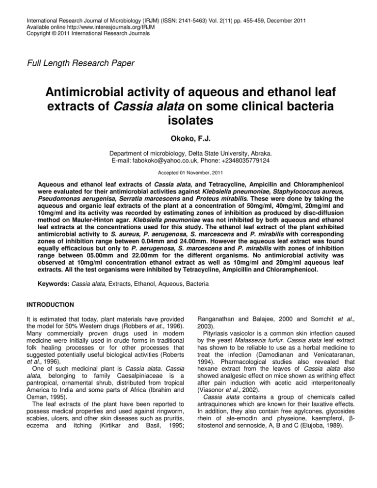
International Research Journal of Microbiology (IRJM) (ISSN: 2141-5463) Vol. 2(11) pp. 455-459, December 2011 Available online http://www.interesjournals.org/IRJM Copyright © 2011 International Research Journals Full Length Research Paper Antimicrobial activity of aqueous and ethanol leaf extracts of Cassia alata on some clinical bacteria isolates Okoko, F.J. Department of microbiology, Delta State University, Abraka. E-mail: fabokoko@yahoo.co.uk, Phone: +2348035779124 Accepted 01 November, 2011 Aqueous and ethanol leaf extracts of Cassia alata, and Tetracycline, Ampicilin and Chloramphenicol were evaluated for their antimicrobial activities against Klebsiella pneumoniae, Staphylococcus aureus, Pseudomonas aerugenisa, Serratia marcescens and Proteus mirabilis. These were done by taking the aqueous and organic leaf extracts of the plant at a concentration of 50mg/ml, 40mg/ml, 20mg/ml and 10mg/ml and its activity was recorded by estimating zones of inhibition as produced by disc-diffusion method on Mauler-Hinton agar. Klebsiella pneumoniae was not inhibited by both aqueous and ethanol leaf extracts at the concentrations used for this study. The ethanol leaf extract of the plant exhibited antimicrobial activity to S. aureus, P. aerugenosa, S. marcescens and P. mirabilis with corresponding zones of inhibition range between 0.04mm and 24.00mm. However the aqueous leaf extract was found equally efficacious but only to P. aerugenosa, S. marcescens and P. mirabilis with zones of inhibition range between 05.00mm and 22.00mm for the different organisms. No antimicrobial activity was observed at 10mg/ml concentration ethanol extract as well as 10mg/ml and 20mg/ml aqueous leaf extracts. All the test organisms were inhibited by Tetracycline, Ampicillin and Chloramphenicol. Keywords: Cassia alata, Extracts, Ethanol, Aqueous, Bacteria INTRODUCTION It is estimated that today, plant materials have provided the model for 50% Western drugs (Robbers et at., 1996). Many commercially proven drugs used in modern medicine were initially used in crude forms in traditional folk healing processes or for other processes that suggested potentially useful biological activities (Roberts et al., 1996). One of such medicinal plant is Cassia alata. Cassia alata, belonging to family Caesalpiniaceae is a pantropical, ornamental shrub, distributed from tropical America to India and some parts of Africa (Ibrahim and Osman, 1995). The leaf extracts of the plant have been reported to possess medical properties and used against ringworm, scabies, ulcers, and other skin diseases such as pruritis, eczema and itching (Kirtikar and Basil, 1995; Ranganathan and Balajee, 2000 and Somchit et al., 2003). Pityriasis vasicolor is a common skin infection caused by the yeast Malassezia furfur. Cassia alata leaf extract has shown to be reliable to use as a herbal medicine to treat the infection (Damodianan and Venicataranan, 1994). Pharmacological studies also revealed that hexane extract from the leaves of Cassia alata also showed analgesic effect on mice shown as writhing effect after pain induction with acetic acid interperitoneally (Viasonor et al., 2002). Cassia alata contains a group of chemicals called antraquinones which are known for their laxative effects. In addition, they also contain free agylcones, glycosides rhein of ale-emodin and physeione, kaempferol, βsitostenol and sennoside, A, B and C (Elujoba, 1989). 456 Int. Res. J. Microbiol. The objective of this study is to investigate the susceptibility of the test bacteria to both ethanol and aqueous extracts of Cassia alata and to compare the efficacy of the extracts with some commercial antibiotics such as Tetracycline, Penicillin and Chloramphenicol. MATERIALS AND METHODS Plant Sample and Source The Cassia alata plant leaves used for this study were obtained from Site 2 of Delta State University, Abraka. They were identified by Dr. S.M. Ayodele and Professor J.M.O. Eze of Botany Department in the same University. Leaves Processing and Extraction Procedure The leaves of Cassia alata were carefully plucked from the stems, washed with distilled water and sun-dried for 4 days after which they were oven-dried for 15 minutes at 40°C to ensure proper drying. The leaves were blended into powder using an electric Osterizer Cycle Blender, Model N0.890-96F, Dallas. One hundred grams (100g) of the blended leaves was weighed into each of the two 500ml conical flasks containing 250ml of distilled water and 250ml of 70% ethanol respectively. The contents were stirred gently using stirring rod. Each flask was corked and properly labeled and kept on a clean table in the laboratory for 72 hours for extraction to take place. After 72 hours of extraction the contents of both flasks were sieved into two sterile 250ml conical flasks using Watman N0.1 filter paper fitted into a glass funnel. The flasks were properly labeled while the supernatants were discarded. The flasks with their contents were transferred into a water bath adjusted to 40°C and left for 72 hours to evaporate the solvent and concentrate the extracts into paste. Collection of Microorganisms Pure cultures of Staphylococcus aureus, Klebsiella pneumoniae, Serratia marcescens, Pseudomonas auriginosa and Proteus mirabilis were obtained from Lahor Research and Medical Center, Benin-City, Edo State on agar slants. They were transferred to the laboratory and identification was based on cultural, morphological and biochemical characteristics according to the Schemes of Buchanan and Gibbons (1974), Cowan and Steel (1974) and Macfaddin (1980). Preparation of Filter Paper Disc Filter paper discs were prepared by cutting paper disc of 3mm in diameter from Watman No.1 filter paper using a punch. The discs were spread out singly on a clean Petridish and placed in an oven at 100°C for 1 hour. They were allowed to cool after which 2ml of the ½, ¼, and 1/8 dilutions of both the aqueous and ethanol extracts were impregnated into different discs using sterile pipettes and labeled accordingly. The discs were left covered for 10minutes to allow the extracts penetrate and soak each disc. Serial Dilution of Extracts Two rows of test tubes consisting of four test tubes per row were set in a test tube rack and rebelled accordingly. Row "A" was to be used for the ethanol extract while row "B" for the aqueous extract dilution. A sterile 5ml pipette was used to dispense 2ml distilled water into each tube in the test tube rack. A fresh sterile pipette was used to transfer 2ml of ethanol extract into the first tube in row "A" and the content of the tube was thoroughly mixed. With a fresh pipette, 2ml of the content of tube 1 was transferred to tube 2 and mixed thoroughly. The same process was repeated for tube 3 and mixed after which 2ml of its content was discarded. Tube 4 serves as control. Similar steps were repeated for the aqueous extract in row 'B'. The final dilutions for both extracts were ½v/v, ¼v/v and 1/8v/v. Preparation of Antibiotics The antibiotics; tetracycline, ampicilin and chloramphenicol of 250mg per capsule were purchased from a local medicine store in Abraka. Each capsule (250mg) was dissolved in 250ml of sterile water and 1ml solution from each capsule was impregnated into different test disc in the Petri dish and labeled accordingly, 1ml of each dilution contains 1mg of the antibiotics. Antibiotic Sensitivity Testing Four different nutrient agar plates were each seeded with the different test organisms. With the aid of a sterile forcep, the impregnated discs were aseptically placed on the agar plates after which the plates were incubate for 24hours at 37°C. The zones of inhibition were measured and recorded in millimeters. Okoko 457 Table 1. Determination of minimum inhibitory concentration of Ethanol and Aqueous extract on indicator bacteria Organisms 80mg/ml Dilutions (Ethanolic) 40mg/ml 20mg/ml 10mg/ml 80mg/ml Dilutions (Aqueous) 40mg/ml 20mg/ml 10mg/ml Klebsiella pneumonia 00.00 00.00 00.00 00.00 00.00 00.00 00.00 00.00 Staphylococcus aureus 11.00 08.00 10.00 00.00 00.00 00.00 00.00 00.00 Pseudomonas aeruginosa 13.00 17.00 12.00 00.00 14.00 11.00 00.00 00.00 Serratia marcescens 24.00 17.00 12.00 00.00 22.00 16.00 05.00 00.00 Proteus mirabilis 23.00 12.00 04.00 00.00 22.00 19.00 00.00 00.00 Table 2. Zones of inhibition (mm) of Cassia alata extract from undiluted aqueous and ethanol extract on test bacteria Microorganisms Klebsiella pneumonia Staphylococcus aureus Pseudomonas aeruginosa Serratia marcescens Proteus mirabilis Determination of Minimum Inhibitory Concentration of Ethanol and Aqueous Extracts of Cassia alata Two fold dilution of both ethanolic and aqueous leaf extracts of Cassia alata was carried out in sterile test tubes to various concentrations of ½v/v, 1/4v/v and 1/8v/v and mixed thoroughly. The set of tubes with the various concentrations were inoculated with the different test organisms at a concentration of 106CFU/ml and incubated at 37°C overnight after which they were examined for visible growth. The least dilution of the extracts that yielded no growth is the minimum inhibitory concentration. RESULTS The results of the antimicrobial determination for the ethanol and aqueous extracts of Cassia alata against the five test organisms were investigated in a disc-diffusion assay. Table 1 shows that Klebsiella is resistant to both ethanol and aqueous extracts of Cassia alata at all Ethanol 00.00 11.00 13.00 24.00 23.00 Aqueous 11.00 00.00 14.00 22.00 22.00 concentrations employed in this study. Staphylococcus aureus, Pseudomonas aeruginosa, Serratia marcescens and Proteus mirabilis were inhibited by the ethanol extract at concentration 20mg/ml – 80mg/ml while showing different zones of inhibition. All organisms were resistant at 10mg/ml ethanol extract concentration showing no zones of inhibition. Among all the test organisms exposed to the ethanolic extract, Serratia marcescens was the most susceptible with 24.00mm zone of inhibition at 80mg/ml concentration. However, a test of the antimicrobial activity of the aqueous extract of the leaves on the test organisms revealed that only Pseudomonas aeruginosa, Serratia marcescens and Proteus mirabilis are susceptible to the extract at the same concentrations as exhibited by the ethanolic extract but uninhibitory to the same organisms at 10mg/ml and 25mg/ml of the extract. Serratia marcescens was found to be slightly susceptible showing 05.00mm zone of inhibition (See Table 1). The antimicrobial activities of the undiluted ethanol and aqueous leaf extracts of Cassia alata is shown in Table 2. Klebsiella pneumoniae was resistant to the ethanol extract but inhibited by the aqueous extract with 458 Int. Res. J. Microbiol. Table 3. Zones of inhibition (mm) of Antibiotics against the indicator bacteria Microorganisms Klebsiella pneumonia Staphylococcus aureus Pseudomonas aeruginosa Serratia marcescens Proteus mirabilis Key: C T A T(1mg/ml) 15.00 13.00 10.00 19.00 15.00 Dilutions C(1mg/ml) 14.00 23.00 08.50 13.00 12.00 A(1mg/ml) 23.00 28.00 12.00 20.00 13.00 Chloramphenicol Tetracycline Ampicilin 11.00mm zone of inhibition while staphylococcus was susceptible to the ethanol extract with 11.00mm zone of inhibition but resistant to the aqueous extract. The other test organisms were inhibited by both extracts showing various zones of inhibition as illustrated in the Table. The effects of the selected antibiotics on the test organisms are shown in Table 3. All the organisms were found to be inhibited by these test antibiotics at the concentration (1mg/ml) used for this study with ampicillin showing the greatest efficacy. DISCUSSION AND CONCLUSION The results obtained from this study shows that the leaves of Cassia alata plant has antimicrobial activities. This observation is not at variance with the work of Somchit et al (2003) who investigated the antimicrobial activities of this plant on several pathogenic microorganisms. Klebsiella pneumoniae was resistant to both extracts used in this study. This could be due to the concentration of the extracts employed in this work or to the chemistry of the cell wall of the organism which may have made it difficult for the active substance to penetrate into the bacteria cells. The other test bacteria were susceptible to both extracts with Serratia marcescens and Proteus mirabilis showing more susceptibility for both extracts. The data obtained as shown in Table 3 showed that Tetracycline, Ampicillin and Chloramphenicol are inhibitory to all test organisms even at low concentrations (1mg/ml) used in this study. The efficacy of the orthodox drugs may be due to the standard method of their production. Most of the bioactive ingredients of the Cassia alata leaf extracts may have been lost due to crude methods of their extraction. The different bioactive components of the leaves have different solubility, hence the ethanolic and aqueous extracts may contain different bioactive substances depending on the solubility of bioactive substances in the two solvents used, This is the cause of the difference in their actions and those of the commercial drugs on the test bacteria. It is clear from the study that the anti microbial activity of the commercial antibiotics is greater than the effects of the plant extracts on the test organisms. The powerful effects of the antibiotics is due to the presence of a mixture of antimicrobial agents present in the antibiotics while plant extracts may lack them or may be present in very low concentration. Tannin is a plant extract which is very sensitive to heat. This calls for a more careful method in the processing of these extracts in order to preserve these delicate ingredients. The results obtained from this study shows that the leaf extracts of Cassia alata has good therapeutic and antimicrobial activities. It is therefore suggested that more studies be carried out to improve on the method of extraction, processing and packaging of extracted products. It could be interesting to also investigate the potentiality of this plant for possible application in foods to increase shelf life or promote safety. REFERENCES Buchanan RE, Gibbons NE (1974). Manual of Determinative th Bacteriology (8 edn). Williams and Wilkens Co. Baltimore, MD. Pp. 126. Cowan ST, Steel KJ (1974) Manual for Identification of Medical Bacteria. (2nd edn) Cambridge University Press, England, pp. 237 Damodianan, Venicatarinan (1994). Study on the Therapeutic Efficacy of Cassia alata Leaf Extract against Pitynasis versicolor. J. Ethnopharmacol.42: 19-23. Elujoba AA, Ajulo OO, Iweobo GO (1989). Chemical and Biological Analysis for Laxative Activity. J. Pharmaceuticals and Biochem. Analysis 7(12): 1453-1457. Ibrahim D, Osman H (1995). Antimicrobial activity of Cassia alata from Okoko 459 Malaysia. J. Ethnopharmacol.45(30): 151-156. Kirtiker, K.R. and Basu, B.D. (1975). Indian Medicinal Plants. Jayyed nd Press Vol.2. 2 Edn. Pp.30-45. Macfaddin FJ (1980). Biochemical Tests for the Identification of Medical Bacteria. (2nd edn). William and Wilkins Company Baltimore pp. 527. Raganathan S, Balajee SA (2000). Antimicrococcus activity of Combination of Cassia alata and Ocimum sanctum. Mycoses; 43: 78, 299-301. Robberts J, Speedle M, Tyier V (1996). Pharmacognosy and Pharmacobiotechnology. Williams and Wilkins Company, Baltimore MD. Pp. 126. Somchit MN, Reezal I, Elycha NI, Mutalih AR (2003). In Vitro Antimicrobial activity of Ethanol and Water Extracts of Cassia alata. J. Ethnopharmacol.84(1): 1-4. Villasenor IM, Canias AP, Pascua MPI, Sabando MN, Soliven LA (2002). Bioactivity Studies on Cassia alata Leaf Extract. Phytother Res. 16: 93-96.
