Anti-AKT1 antibody ab59284 Product datasheet 2 Images Overview
advertisement
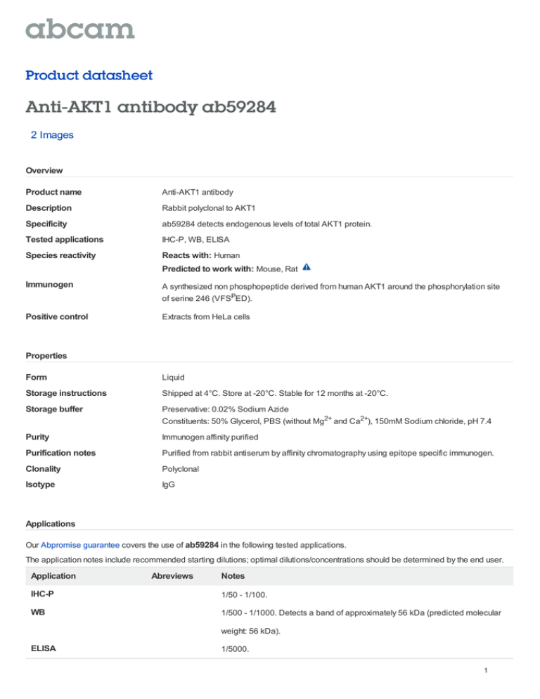
Product datasheet Anti-AKT1 antibody ab59284 2 Images Overview Product name Anti-AKT1 antibody Description Rabbit polyclonal to AKT1 Specificity ab59284 detects endogenous levels of total AKT1 protein. Tested applications IHC-P, WB, ELISA Species reactivity Reacts with: Human Predicted to work with: Mouse, Rat Immunogen A synthesized non phosphopeptide derived from human AKT1 around the phosphorylation site of serine 246 (VFSPED). Positive control Extracts from HeLa cells Properties Form Liquid Storage instructions Shipped at 4°C. Store at -20°C. Stable for 12 months at -20°C. Storage buffer Preservative: 0.02% Sodium Azide Constituents: 50% Glycerol, PBS (without Mg2+ and Ca2+), 150mM Sodium chloride, pH 7.4 Purity Immunogen affinity purified Purification notes Purified from rabbit antiserum by affinity chromatography using epitope specific immunogen. Clonality Polyclonal Isotype IgG Applications Our Abpromise guarantee covers the use of ab59284 in the following tested applications. The application notes include recommended starting dilutions; optimal dilutions/concentrations should be determined by the end user. Application Abreviews Notes IHC-P 1/50 - 1/100. WB 1/500 - 1/1000. Detects a band of approximately 56 kDa (predicted molecular weight: 56 kDa). ELISA 1/5000. 1 Target Function Plays a role as a key modulator of the AKT-mTOR signaling pathway controlling the tempo of the process of newborn neurons integration during adult neurogenesis, including correct neuron positioning, dendritic development and synapse formation (By similarity). General protein kinase capable of phosphorylating several known proteins. Phosphorylates TBC1D4. Signals downstream of phosphatidylinositol 3-kinase (PI(3)K) to mediate the effects of various growth factors such as platelet-derived growth factor (PDGF), epidermal growth factor (EGF), insulin and insulin-like growth factor I (IGF-I). Plays a role in glucose transport by mediating insulininduced translocation of the GLUT4 glucose transporter to the cell surface. Mediates the antiapoptotic effects of IGF-I. Mediates insulin-stimulated protein synthesis by phosphorylating TSC2 at 'Ser-939' and 'Thr-1462', thereby activating mTORC1 signaling and leading to both phosphorylation of 4E-BP1 and in activation of RPS6KB1. Promotes glycogen synthesis by mediating the insulin-induced activation of glycogen synthase. The activated form can suppress FoxO gene transcription and promote cell cycle progression. Essential for the SPATA13mediated regulation of cell migration and adhesion assembly and disassembly. Tissue specificity Expressed in all human cell types so far analyzed. The Tyr-176 phosphorylated form shows a significant increase in expression in breast cancers during the progressive stages i.e. normal to hyperplasia (ADH), ductal carcinoma in situ (DCIS), invasive ductal carcinoma (IDC) and lymph node metastatic (LNMM) stages. Involvement in disease Defects in AKT1 are a cause of susceptibility to breast cancer (BC) [MIM:114480]. A common malignancy originating from breast epithelial tissue. Breast neoplasms can be distinguished by their histologic pattern. Invasive ductal carcinoma is by far the most common type. Breast cancer is etiologically and genetically heterogeneous. Important genetic factors have been indicated by familial occurrence and bilateral involvement. Mutations at more than one locus can be involved in different families or even in the same case. Defects in AKT1 are associated with colorectal cancer (CRC) [MIM:114500]. Defects in AKT1 are associated with susceptibility to ovarian cancer [MIM:604370]; also called susceptibility to familial breast-ovarian cancer type 1 (BROVCA1). Sequence similarities Belongs to the protein kinase superfamily. AGC Ser/Thr protein kinase family. RAC subfamily. Contains 1 AGC-kinase C-terminal domain. Contains 1 PH domain. Contains 1 protein kinase domain. Domain Binding of the PH domain to the phosphatidylinositol 3-kinase alpha (PI(3)K) results in its targeting to the plasma membrane. The PH domain mediates interaction with TNK2 and Tyr-176 is also essential for this interaction. The AGC-kinase C-terminal mediates interaction with THEM4. Post-translational modifications Phosphorylation on Thr-308, Ser-473 and Tyr-474 is required for full activity. Activated TNK2 phosphorylates it on Tyr-176 resulting in its binding to the anionic plasma membrane phospholipid PA. This phosphorylated form localizes to the cell membrane, where it is targeted by PDPK1 and PDPK2 for further phosphorylations on Thr-308 and Ser-473 leading to its activation. Ser-473 phosphorylation by mTORC2 favors Thr-308 phosphorylation by PDPK1. Ser-473 phosphorylation is enhanced by interaction with AGAP2 isoform 2 (PIKE-A). Ser-473 phosphorylation is enhanced in focal cortical dysplasias with Taylor-type balloon cells. Ubiquitinated; undergoes both 'Lys-48'- and 'Lys-63'-linked polyubiquitination. TRAF6-induced 'Lys-63'-linked AKT1 ubiquitination is critical for phosphorylation and activation. When ubiquitinated, it translocates to the plasma membrane, where it becomes phosphorylated. When fully phosphorylated and translocated into the nucleus, undergoes 'Lys-48'-polyubiquitination catalyzed by TTC3, leading to its degradation by the proteasome. Cellular localization Cytoplasm. Nucleus. Cell membrane. Nucleus after activation by integrin-linked protein kinase 1 (ILK1). Nuclear translocation is enhanced by interaction with TCL1A. Phosphorylation on Tyr-176 by TNK2 results in its localization to the cell membrane where it is targeted for further 2 phosphorylations on Thr-308 and Ser-473 leading to its activation and the activated form translocates to the nucleus. Anti-AKT1 antibody images All lanes : Anti-AKT1 antibody (ab59284) at 1/500 dilution Lane 1 : Extracts from HeLa cells, treated with Etoposide (25uM, 24hours) with no immunizing peptide Lane 2 : Extracts from HeLa cells, treated with Etoposide (25uM, 24hours) with immunizing peptide Western blot - AKT1 antibody (ab59284) Predicted band size : 56 kDa Immunohistochemistry analysis of paraffinembedded human ovary tissue, using ab59284 at 1/50 dilution. The picture on the right is blocked with the synthesized peptide. Immunohistochemistry (Formalin/PFA-fixed paraffin-embedded sections) - Anti-AKT1 antibody (ab59284) Please note: All products are "FOR RESEARCH USE ONLY AND ARE NOT INTENDED FOR DIAGNOSTIC OR THERAPEUTIC USE" Our Abpromise to you: Quality guaranteed and expert technical support Replacement or refund for products not performing as stated on the datasheet Valid for 12 months from date of delivery Response to your inquiry within 24 hours We provide support in Chinese, English, French, German, Japanese and Spanish Extensive multi-media technical resources to help you We investigate all quality concerns to ensure our products perform to the highest standards If the product does not perform as described on this datasheet, we will offer a refund or replacement. For full details of the Abpromise, please visit http://www.abcam.com/abpromise or contact our technical team. Terms and conditions Guarantee only valid for products bought direct from Abcam or one of our authorized distributors 3 4
