To see what the sfg1 and sfg2 do we can... over express these proteins. We can clone the sfg1...
advertisement
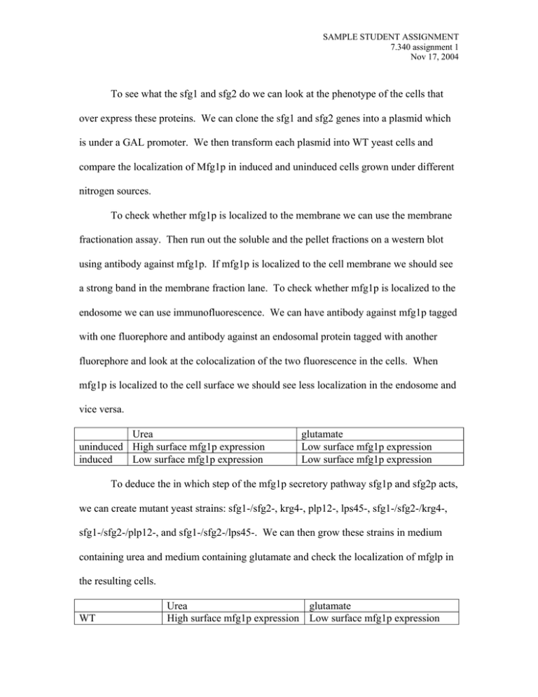
SAMPLE STUDENT ASSIGNMENT 7.340 assignment 1 Nov 17, 2004 To see what the sfg1 and sfg2 do we can look at the phenotype of the cells that over express these proteins. We can clone the sfg1 and sfg2 genes into a plasmid which is under a GAL promoter. We then transform each plasmid into WT yeast cells and compare the localization of Mfg1p in induced and uninduced cells grown under different nitrogen sources. To check whether mfg1p is localized to the membrane we can use the membrane fractionation assay. Then run out the soluble and the pellet fractions on a western blot using antibody against mfg1p. If mfg1p is localized to the cell membrane we should see a strong band in the membrane fraction lane. To check whether mfg1p is localized to the endosome we can use immunofluorescence. We can have antibody against mfg1p tagged with one fluorephore and antibody against an endosomal protein tagged with another fluorephore and look at the colocalization of the two fluorescence in the cells. When mfg1p is localized to the cell surface we should see less localization in the endosome and vice versa. Urea uninduced High surface mfg1p expression induced Low surface mfg1p expression glutamate Low surface mfg1p expression Low surface mfg1p expression To deduce the in which step of the mfg1p secretory pathway sfg1p and sfg2p acts, we can create mutant yeast strains: sfg1-/sfg2-, krg4-, plp12-, lps45-, sfg1-/sfg2-/krg4-, sfg1-/sfg2-/plp12-, and sfg1-/sfg2-/lps45-. We can then grow these strains in medium containing urea and medium containing glutamate and check the localization of mfglp in the resulting cells. WT Urea glutamate High surface mfg1p expression Low surface mfg1p expression SAMPLE STUDENT ASSIGNMENT 7.340 assignment 1 Nov 17, 2004 Sfg1-/sfg2Krg4Plp12Lps45Sfg1-/sfg2-/plp1Sfg1-/sfg2-/lps45Sfg1-/sfg2-/krg4- High surface mfg1p expression Low surface mfg1p expression Low surface mfg1p expression Low surface mfg1p expression Low surface mfg1p expression Low surface mfg1p expression High surface mfg1p expression High surface mfg1p expression Low surface mfg1p expression Low surface mfg1p expression Low surface mfg1p expression Low surface mfg1p expression Low surface mfg1p expression High surface mfg1p expression From this data we can see that sfg1p/sfg2p must act on mfg1p before krg4 and after plp12- and lps45-. Since we already know that plp12 and lps45 are involved in vesicular transport from the golgi to the endosome, and that krg4 is involved in the recycling of mfg1p from the endosome back to the golgi, we know that sfg1p/sfg2p must act somewhere in the cycling of mfg1p between the golgi and the endosome. To check the role of mrt5p in the secretory pathway of mfg1p we can create a mrt5-1 mutant and compare the ubiquitination of mfg1p in WT and mutant yeast cells grown under either urea or glutamate. We will use immunoprecipitation to isolate mfg1p from cell lysate, and then run out the precipitate on a western blot using antibody against ubiquitin. In WT cells grown under both urea and glutamate we should see a band because mfg1p is ubiquitinated no matter what the nitrogen source is, but in the mutant cell we will not see a band because without mrt5p ubiquitin will not be able to attach to mfg1p. This observation should not change with the change of nitrogen source either because without an E3 ligase mfg1p cannot get ubiquitinated no matter what the nitrogen source is. So we can deduce that mrt5p is probably the E3 ligase. To see whether sfg1 and sfg2 are involved in the ubiquitination of mfg1p we can check its interaction with the E3 ligase, Mrt5p. We can use a immunoprecipitation experiment using antibody against mrt5p. Then we can run out the precipitated protein on a western blot using antibodies against sfg1 and sfg2. In cells grown under glutamate SAMPLE STUDENT ASSIGNMENT 7.340 assignment 1 Nov 17, 2004 we should see a band showing up because in these cells sfg1p and sfg2p interacts with mrt5p, affecting the ubiquitination of mfg1p, which allows it to be localized to the endosome and degraded. In cells grown under urea we will not see a band showing up because in these cells sfg1p and sfg2p are not interacting with mrt5p, which allows mfglp to be sorted to the cell surface. To check how sfg1p and sfg2p affect the ubiquitination of mfg1p we can compare the mfg1p ubiquitination of WT cells and sfg1-/sfg2- cells grown under glutamate. We can use immunoprecipitation with antibody against mfg1p and then run out the precipitates on a westernblot detecting with antibody against ubiquitin. In sfg1-/sfg2cells we will see lower molecular weight bands relative to the WT because without sfg1 and sfg2 mrt5p can only monoubquitinate mfg1p. Where as in WT cells we will see higher molecular weight bands because mfg1p is polyubiquitinated by the sfg1/sfg2/mrt5 complex. To check the effect of ubiquitination on the localization of mfg1p we can feed the cells with a ubiquitin analog that cannot polymerize so mfg1p can only be monoubiquitinated. Growing these cells under urea we will see mfg1p localize to the cell membrane because they are monoubiquitinated. However when these cells are grown under glutamate we will also see the same localization at the cell membrane because although the sfg1/sfg2/mrt5 complex is active in trying to polyubiquitinate mfg1p the ubiquitin cannot polymerize, thus the mfg1p will still end up at the cell surface. These results tell us that mfg1p is sorted according to its ubiquidination status.
