Document 13310579
advertisement
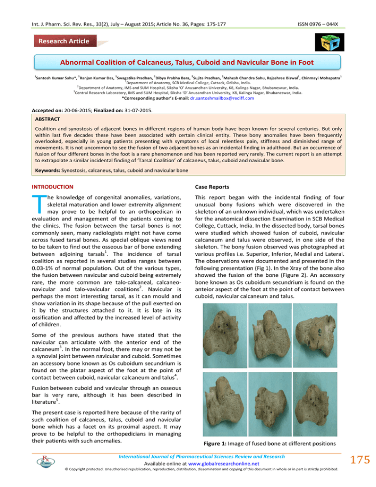
Int. J. Pharm. Sci. Rev. Res., 33(2), July – August 2015; Article No. 36, Pages: 175-177 ISSN 0976 – 044X Research Article Abnormal Coalition of Calcaneus, Talus, Cuboid and Navicular Bone in Foot 1 1 1 1 2 3 1 1 Santosh Kumar Sahu*, Ranjan Kumar Das, Swagatika Pradhan, Dibya Prabha Bara, Sujita Pradhan, Mahesh Chandra Sahu, Rajashree Biswal , Chinmayi Mohapatra 1 Department of Anatomy, SCB Medical College, Cuttack, Odisha, India. 2 Department of Anatomy, IMS and SUM Hospital, Siksha ‘O’ Anusandhan University, K8, Kalinga Nagar, Bhubaneswar, India. 3 Central Research Laboratory, IMS and SUM Hospital, Siksha ‘O’ Anusandhan University, K8, Kalinga Nagar, Bhubaneswar, India. *Corresponding author’s E-mail: dr.santoshmailbox@rediff.com Accepted on: 20-06-2015; Finalized on: 31-07-2015. ABSTRACT Coalition and synostosis of adjacent bones in different regions of human body have been known for several centuries. But only within last five decades these have been associated with certain clinical entity. These bony anomalies have been frequently overlooked, especially in young patients presenting with symptoms of local relentless pain, stiffness and diminished range of movements. It is not uncommon to see the fusion of two adjacent bones as an incidental finding in adulthood. But an occurrence of fusion of four different bones in the foot is a rare phenomenon and has been reported very rarely. The current report is an attempt to extrapolate a similar incidental finding of ‘Tarsal Coalition’ of calcaneus, talus, cuboid and navicular bone. Keywords: Synostosis, calcaneus, talus, cuboid and navicular bone INTRODUCTION Case Reports T he knowledge of congenital anomalies, variations, skeletal maturation and lower extremity alignment may prove to be helpful to an orthopedican in evaluation and management of the patients coming to the clinics. The fusion between the tarsal bones is not commonly seen, many radiologists might not have come across fused tarsal bones. As special oblique views need to be taken to find out the osseous bar of bone extending between adjoining tarsals1. The incidence of tarsal coalition as reported in several studies ranges between 0.03-1% of normal population. Out of the various types, the fusion between navicular and cuboid being extremely rare, the more common are talo-calcaneal, calcaneonavicular and talo-vavicular coalitions2. Navicular is perhaps the most interesting tarsal, as it can mould and show variation in its shape because of the pull exerted on it by the structures attached to it. It is late in its ossification and affected by the increased level of activity of children. This report began with the incidental finding of four unusual bony fusions which were discovered in the skeleton of an unknown individual, which was undertaken for the anatomical dissection Examination in SCB Medical College, Cuttack, India. In the dissected body, tarsal bones were studied which showed fusion of cuboid, navicular calcaneum and talus were observed, in one side of the skeleton. The bony fusion observed was photographed at various profiles i.e. Superior, Inferior, Medial and Lateral. The observations were documented and presented in the following presentation (Fig 1). In the Xray of the bone also showed the fusion of the bone (Figure 2). An accessory bone known as Os cuboidum secundrium is found on the anteior aspect of the foot at the point of contact between cuboid, navicular calcaneum and talus. Some of the previous authors have stated that the navicular can articulate with the anterior end of the calcaneum3. In the normal foot, there may or may not be a synovial joint between navicular and cuboid. Sometimes an accessory bone known as Os cuboidum secundrium is found on the platar aspect of the foot at the point of contact between cuboid, navicular calcaneum and talus4. Fusion between cuboid and vavicular through an osseous bar is very rare, although it has been described in 5 literature . The present case is reported here because of the rarity of such coalition of calcaneus, talus, cuboid and navicular bone which has a facet on its proximal aspect. It may prove to be helpful to the orthopedicians in managing their patients with such anomalies. Figure 1: Image of fused bone at different positions International Journal of Pharmaceutical Sciences Review and Research Available online at www.globalresearchonline.net © Copyright protected. Unauthorised republication, reproduction, distribution, dissemination and copying of this document in whole or in part is strictly prohibited. 175 © Copyright pro Int. J. Pharm. Sci. Rev. Res., 33(2), July – August 2015; Article No. 36, Pages: 175-177 ISSN 0976 – 044X disturbed. These abnormal movements increases stress on other joints of foot leading to wear and tear of joint eventually causing pain. Also, muscles in relation to these joints undergo spasm due to stress of abnormal movements leading to pain and restriction of movement of limb. Sanghyeok, classified talocalcaneal coalition into four different types by using CT and MRI, these are: Type I- Linear with or without posterior hooking Type II- Talar overgrowth Type III- Calcaneal overgrowth Type IV14 Complete Osseous. But, the present report was done in loose skeleton of an unknown person whose relevant case history or clinical findings were not available. Thus, the findings cannot be further studied using any radiological modalities of investigation. Findings on observation show a more or less Type IV coalition in the bones. Figure 2: X ray of the fused bone DISCUSSION In Fused tarsal bone, the fusion may be either congenital or acquired6. It is difficult to identify the case of tarsal synostosis, whether it is congenital or acquired without sufficient clinical data and case history to support the findings. Tarsal fusion anomalies are likely to be associated with disturbance of Pax-1 gene expression in the developing foot7. Acquired tarsal synostosis is generally associated with diseases like tuberculosis, other infections, juvenile rheumatoid arthritis and trauma. All these abnormalities may lead to clinical signs and symptoms which are: osseous malformation (synostosis), and signs of peripheral nerve irritation such as pain, burning sensations and cramps, signs of nerve compression such as hyperaesthesia / anaesthesia, weakness/paralysis8. Coalition of the tarsal bones has been known for several centuries. But it is often overlooked as a cause of foot pain in patients presenting with complaint of non-traumatic foot pain. Baffon was probably the first to recognize the tarsal coalition, although the undated specimen in museum of Royal College of Surgeons of England described by John Hunter probably dated from 1760-1770.9 The talo-calcaneal fusion was first described.10 It was described coalitions as fibrous (syndesmosis), cartilaginous (synchondrosis), and osseous (synostosis).11 The most common types of coalitions are calcaneonavicular, talonavicular, calcaneocuboidal, cubonavicular and talocalcaneal. Incidence of talocalcaneal fusion is <1% to 1%. In the same year proposed the theory of impaired segmentation and undifferentiating of primordial mesenchymal tissue which resulted in coalitions.12 One another study described the congenital type as an extension of an accessory ossicle into adjacent tarsal bones. Tarsal 13 Coalition is mostly a congenital condition. Most eventfully it is inherited as a disorder of autosomal dominant inheritance. It occurs due to the failure of segmentation of mesenchyme with the absence of normal joint formation. It becomes a problem since it restricts the movements of other joints of foot which are CONCLUSION The fusion between the tarsal bones may cause pain and stiffness in foot especially in case of children with congenital flat foot, after the ossification of the tarsal bones are complete, which might be pronounced by injury. So, while considering the causes of flat foot, orthopedicians must keep in mind such coalition as one of the differential diagnosis. REFERENCES 1. Chambers CH, Congenital anomalies of the tarsal Navicular with particular reference to calcaneonavicular coalition, Br J Radiol, 23(274), 1950, 306-308. 2. Feliu EC, Cubo-navicular synostosis-A case report Acta Orthopaedica belgica, 57(3), 1991, 306-308. 3. Trolle D, Accessory bones of the human foot: A radiological, histoembryological, comparatice anatomical and genetic study, Musksgaard Copenhagen, 1948, 232254. 4. Trolle D, Accessory bones of the human foot: A radiological, histoembryological, comparative anatomical and genetic stusy, Musksgaard Copenhagen, 1948, 232254. 5. Burman MS, Lapidus PW, The functional disturbances caused by the inconstant bones and sesamoids of the foot Arch Surg, 22(6), 1931, 936-975. 6. Erdil H, Yidiz N, Cimen M, Congenital Fusion of Cervical Vertebrae and its clinical significance, Journal of Anatomical Society of India, 52, 2003, 125-27. 7. David KM, Coop AJ, Stevens JM, Hayward RD and Crockard HA, Split cervical spinal cord with Klippe- Feil syndrome: Seven Cases, Journal of Neurology, 119(6), 1996, 1859-72. 8. Tiwari A, Chandra N, Naresh M, Pandey A and Tiwari K. Congenital Abnormal Cervical Vertebrae - A Case Report. Journal of Anatomical Society of India, 51, 2002, 68-9. 9. Buffon GLL, Comte de. Histoire Naturelle, Generale et Particulire (Panckoucke) Paris 3 47. Cave AJE. Proceedings Anatomical Society. Journal of Anatomical Society of London, 72, 1769, 319. International Journal of Pharmaceutical Sciences Review and Research Available online at www.globalresearchonline.net © Copyright protected. Unauthorised republication, reproduction, distribution, dissemination and copying of this document in whole or in part is strictly prohibited. 176 © Copyright pro Int. J. Pharm. Sci. Rev. Res., 33(2), July – August 2015; Article No. 36, Pages: 175-177 10. Zuckerkandl E, Ueber einen Fall von Synostose zwischen Talus und Calcaneus, Allgemeine Weiner Mediinische Zeitung, 22, 1877, 293-294. 11. Buckholtz JM. Baltimore, Peroneal spastic flatfoot, Fundamentals of Foot Surgery, 51, 68-9. 12. LeBoucq H. De la, sondure congenitale de certains os du tarse. Bulletin of a Royal Academy of Medicine in Belgium, 4, 1896, 103. ISSN 0976 – 044X 13. Pfitzner W. Die variationem im aufbau des fusskelets. Morphologisches Arbeiten, 6, 1896, 245-527. 14. Sanghyeok Lim, Hyeon Kyeong Lee, Sooho Bae, Nae-jung Rim and Jaeho Cho (). A radiological classification system for talocalcaneal coalition based on a multi-planar imaging study using CT and MRI, Insights Imaging, 4, 2013, 563– 567. Source of Support: Nil, Conflict of Interest: None. International Journal of Pharmaceutical Sciences Review and Research Available online at www.globalresearchonline.net © Copyright protected. Unauthorised republication, reproduction, distribution, dissemination and copying of this document in whole or in part is strictly prohibited. 177 © Copyright pro

