Document 13309700
advertisement
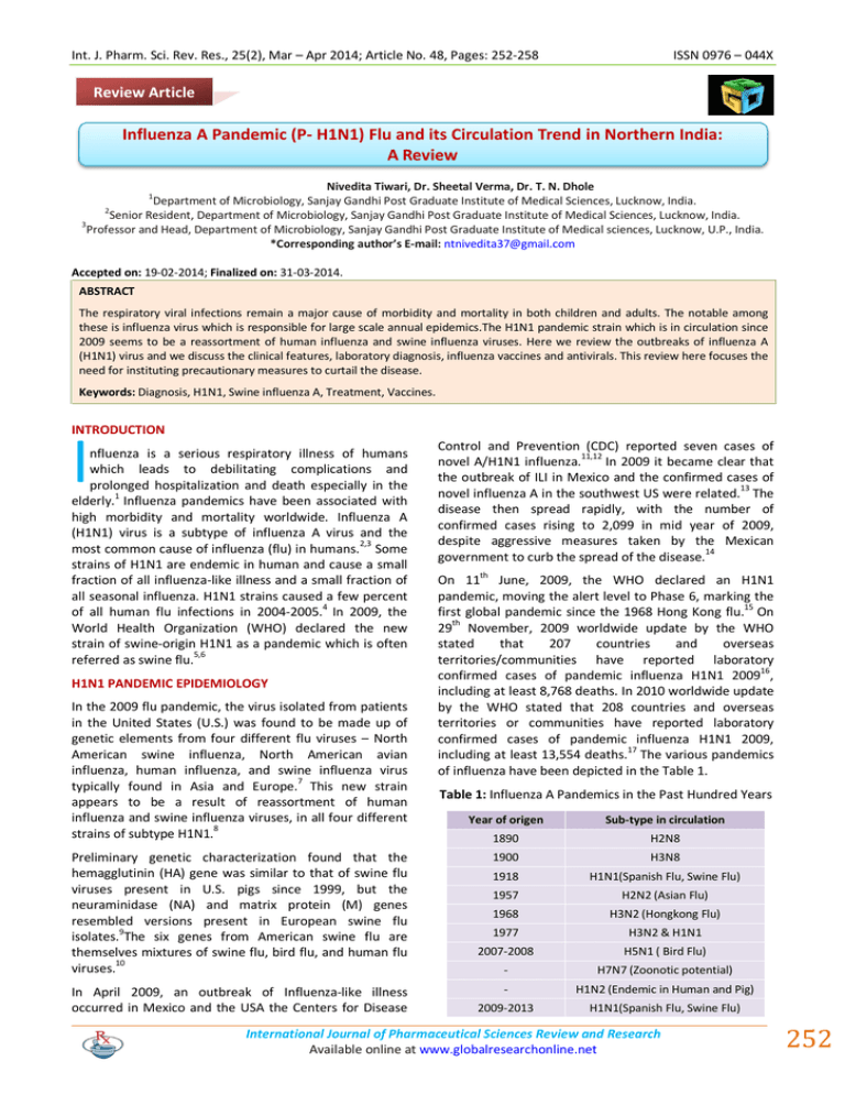
Int. J. Pharm. Sci. Rev. Res., 25(2), Mar – Apr 2014; Article No. 48, Pages: 252-258 ISSN 0976 – 044X Review Article Influenza A Pandemic (P- H1N1) Flu and its Circulation Trend in Northern India: A Review Nivedita Tiwari, Dr. Sheetal Verma, Dr. T. N. Dhole Department of Microbiology, Sanjay Gandhi Post Graduate Institute of Medical Sciences, Lucknow, India. 2 Senior Resident, Department of Microbiology, Sanjay Gandhi Post Graduate Institute of Medical Sciences, Lucknow, India. 3 Professor and Head, Department of Microbiology, Sanjay Gandhi Post Graduate Institute of Medical sciences, Lucknow, U.P., India. *Corresponding author’s E-mail: ntnivedita37@gmail.com 1 Accepted on: 19-02-2014; Finalized on: 31-03-2014. ABSTRACT The respiratory viral infections remain a major cause of morbidity and mortality in both children and adults. The notable among these is influenza virus which is responsible for large scale annual epidemics.The H1N1 pandemic strain which is in circulation since 2009 seems to be a reassortment of human influenza and swine influenza viruses. Here we review the outbreaks of influenza A (H1N1) virus and we discuss the clinical features, laboratory diagnosis, influenza vaccines and antivirals. This review here focuses the need for instituting precautionary measures to curtail the disease. Keywords: Diagnosis, H1N1, Swine influenza A, Treatment, Vaccines. INTRODUCTION I nfluenza is a serious respiratory illness of humans which leads to debilitating complications and prolonged hospitalization and death especially in the elderly.1 Influenza pandemics have been associated with high morbidity and mortality worldwide. Influenza A (H1N1) virus is a subtype of influenza A virus and the most common cause of influenza (flu) in humans.2,3 Some strains of H1N1 are endemic in human and cause a small fraction of all influenza-like illness and a small fraction of all seasonal influenza. H1N1 strains caused a few percent of all human flu infections in 2004-2005.4 In 2009, the World Health Organization (WHO) declared the new strain of swine-origin H1N1 as a pandemic which is often referred as swine flu.5,6 H1N1 PANDEMIC EPIDEMIOLOGY In the 2009 flu pandemic, the virus isolated from patients in the United States (U.S.) was found to be made up of genetic elements from four different flu viruses – North American swine influenza, North American avian influenza, human influenza, and swine influenza virus 7 typically found in Asia and Europe. This new strain appears to be a result of reassortment of human influenza and swine influenza viruses, in all four different 8 strains of subtype H1N1. Preliminary genetic characterization found that the hemagglutinin (HA) gene was similar to that of swine flu viruses present in U.S. pigs since 1999, but the neuraminidase (NA) and matrix protein (M) genes resembled versions present in European swine flu isolates.9The six genes from American swine flu are themselves mixtures of swine flu, bird flu, and human flu viruses.10 In April 2009, an outbreak of Influenza-like illness occurred in Mexico and the USA the Centers for Disease Control and Prevention (CDC) reported seven cases of novel A/H1N1 influenza.11,12 In 2009 it became clear that the outbreak of ILI in Mexico and the confirmed cases of novel influenza A in the southwest US were related.13 The disease then spread rapidly, with the number of confirmed cases rising to 2,099 in mid year of 2009, despite aggressive measures taken by the Mexican government to curb the spread of the disease.14 On 11th June, 2009, the WHO declared an H1N1 pandemic, moving the alert level to Phase 6, marking the first global pandemic since the 1968 Hong Kong flu.15 On 29th November, 2009 worldwide update by the WHO stated that 207 countries and overseas territories/communities have reported laboratory confirmed cases of pandemic influenza H1N1 200916, including at least 8,768 deaths. In 2010 worldwide update by the WHO stated that 208 countries and overseas territories or communities have reported laboratory confirmed cases of pandemic influenza H1N1 2009, including at least 13,554 deaths.17 The various pandemics of influenza have been depicted in the Table 1. Table 1: Influenza A Pandemics in the Past Hundred Years Year of origen Sub-type in circulation 1890 H2N8 1900 H3N8 1918 H1N1(Spanish Flu, Swine Flu) 1957 H2N2 (Asian Flu) 1968 H3N2 (Hongkong Flu) 1977 H3N2 & H1N1 2007-2008 H5N1 ( Bird Flu) - H7N7 (Zoonotic potential) - H1N2 (Endemic in Human and Pig) 2009-2013 H1N1(Spanish Flu, Swine Flu) International Journal of Pharmaceutical Sciences Review and Research Available online at www.globalresearchonline.net 252 Int. J. Pharm. Sci. Rev. Res., 25(2), Mar – Apr 2014; Article No. 48, Pages: 252-258 ISSN 0976 – 044X Transmission Adults and adolescents Person-to-person transmission The most common clinical findings of the 2009 H1N1 influenza A pandemic have been fever, cough, sore throat, malaise, and headache, vomiting and diarrhea have also been common, both of which are unusual features of seasonal influenza.29 Other frequent findings have included chills, myalgias, and arthralgias. Rhabdomyolysis has been reported rarely.30 Influenza virus can be transmitted through sneezing and 18 coughing via large-particle droplets. In addition to respiratory secretions, certain other body fluids (e.g. diarrheal stool) should also be considered potentially infectious. In contrast to previous outbreaks of swine influenza viruses, the pandemic of H1N1 influenza A infection that began in March 2009 appears to involve sustained human-to-human transmission, as suggested by the large numbers of patients with respiratory illnesses identified within a short period of time at various 19 locations around the world. Several of the isolates causing disease in the U.S. have been found to be nearly genetically identical to isolates in Mexico, supportive of 20 person-to-person transmission. Viral shedding Children Young children are less likely to have the usual influenza signs and symptoms, such as fever and cough. Infants may present with fever and lethargy, and may not have 31 cough or other respiratory symptoms. Symptoms of severe disease in infants and young children may include apnea, tachypnea, dyspnea, cyanosis, dehydration, 32 altered mental status, and extreme irritability. Definitions of illness severity Influenza shedding begins the day prior to symptom onset and can persist for five to seven days in immunocompetent individuals.21 Longer periods of shedding may occur in children (especially young infants), elderly adults, patients with chronic illnesses, and immunocompromised hosts.22 The amount of virus shed is greatest during the first two to three days of illness.23 Although it is thought that immunocompetent patients with pandemic H1N1 influenza A virus infection are likely to be contagious from one day prior to the development of signs and symptoms until resolution of fever, longer periods of shedding were also detected several cases.24 Secondary attack rates There are conflicting data regarding whether secondary attack rates are higher with H1N1 influenza infection compared to seasonal influenza.25 Environmental transmission Transmission via contact with surfaces that have been contaminated with respiratory droplets or by aerosolized small-particle droplets may also occur.26 Role of pigs Pigs play an important role in interspecies transmission of influenza virus. Susceptible pig cells possess receptors for both avian (alpha 2-3-linked sialic acids) and human influenza strains (alpha 2-6-linked sialic acids), which allow reassortment of influenza virus genes from different species if a pig cell is infected with more than one strain.27 Clinical Manifestations The signs and symptoms of influenza caused by pandemic H1N1 influenza A virus are similar to those of seasonal influenza, although gastrointestinal manifestations (vomiting) appear to be more common with pandemic H1N1 influenza A.28 The U.S. CDC has categorized the severity of illness from influenza as follows;33 A. Mild or uncomplicated illness is characterized by fever, cough, sore throat, rhinorrhea, muscle pain, headache, chills, malaise, and sometimes diarrhea and vomiting, but no shortness of breath and little change in chronic health conditions. B. Progressive illness is characterized by typical symptoms plus signs or symptoms such as chest pain, poor oxygenation (e.g. tachypnea, hypoxia, labored breathing in children), cardiopulmonary insufficiency (e.g. hypotension), CNS impairment (e.g. confusion, altered mental status), severe dehydration, or exacerbations of chronic conditions (e.g. asthma, chronic obstructive pulmonary disease, chronic renal failure, diabetes or other cardiovascular conditions). C. Severe or complicated illness is characterized by signs of lower respiratory tract disease (e.g. hypoxia requiring supplemental oxygen, abnormal chest radiograph, mechanical ventilation), CNS findings (encephalitis, encephalopathy), complications of hypotension (shock, organ failure), myocarditis or rhabdomyolisis, or invasive secondary bacterial infection based on laboratory testing or clinical signs (e.g. persistent high fever and other symptoms beyond three days). Laboratory Diagnosis Collection, storage and transport of specimen Clinical specimens that are to be tested for influenza viruses can be collected either as part of routine patient care (through sentinel surveillance) or during outbreak investigations.34 The successful isolation of an influenza virus depends upon the prompt collection of high-quality specimens, the rapid transportation of specimens to the testing laboratory, and appropriate transport and storage conditions prior to testing. Ideally, respiratory specimens and acute-phase serological specimens should be International Journal of Pharmaceutical Sciences Review and Research Available online at www.globalresearchonline.net 253 Int. J. Pharm. Sci. Rev. Res., 25(2), Mar – Apr 2014; Article No. 48, Pages: 252-258 collected within 3 days of the onset of clinical symptoms.35, 36 Processing of clinical specimens for virus isolation Virus isolation is a highly sensitive and very useful 37 procedure for the diagnosis of viral infection. One important advantage of virus isolation is that it amplifies the amount of virus in the original specimen, thus producing a sufficient quantity for further antigenic and genetic characterization, and for drug susceptibility 38 testing if required. Virus isolation in cell culture The isolation of viruses in cell cultures is increasingly becoming the gold standard for virus diagnosis. However a laboratory must maintain several cell lines to allow for the detection of a variety of respiratory pathogens. Since standard virus-isolation procedures take several days before results are available they are usually of limited use 39 in clinical settings for the prompt diagnosis of influenza. ISSN 0976 – 044X many epidemiological and immunological studies and for evaluation of the antibody response following vaccination. Serological methods are also very useful in situations where identification of the virus is not feasible (e.g. after viral shedding has stopped). Demonstration of an acute influenza infection using serology requires a significant increase in antibody titres (i.e. 4-fold or greater) between acute-phase and convalescent phase serum samples. The demonstration of such a significant increase may establish the diagnosis of a recent infection even when attempts to detect the virus are negative. Serological diagnosis of influenza by microneutralization assay In recent years, the use of cell cultures has surpassed the use of embryonated eggs to isolate and culture influenza viruses. However, only viruses grown in embryonated eggs can be used as seed viruses for the production of the majority of influenza vaccines. For this reason, laboratories that have the capability to isolate influenza viruses in eggs are encouraged to maintain this capacity.40 Serological methods such as the HAI test rarely yield an early diagnosis of acute influenza virus infection. Although conventional neutralization tests for influenza viruses (based on the inhibition of cytopathogenic effect formation in MDCK cell culture) are laborious and rather slow, a microneutralization assay using microtitre plates in combination with an ELISA to detect virus-infected cells can yield results within two days. The microneutralization assay is a highly sensitive and specific assay for detecting virus-specific neutralizing antibodies to influenza viruses in human and animal sera, potentially including the detection of human antibodies to avian subtypes. Testing can be carried out quickly once a novel virus is identified and often before purified viral proteins become available for use in other assays.41 Identification of the haemagglutinin subtype of viral isolates by haemagglutination inhibition testing Identification of neuraminidase subtype by neuraminidase assay and neuraminidase inhibition test The haemagglutination inhibition (HAI) test is an extremely reliable assay for typing, subtyping and further determining the antigenic characteristics of influenza viral isolates provided that the reference antisera used contain antibodies to currently circulating viruses. The antisera used are based on antigen preparations derived from either the wildtype strain or a high-growth reassortant made using the wild-type strain or an antigenically equivalent strain. The disadvantages of the HAI test include the need to remove nonspecific inhibitors of haemagglutination that occur naturally in sera; the need to standardize reference and test antigens each time a test is performed; and the need for specialized expertise in reading the results of the test. Nevertheless, the HAI test remains the assay of choice for global influenza surveillance and for determining the antigenic characteristics of influenza viral isolates. There are two basic forms of assay for influenza virus neuraminidase (NA) based on the use of different substrate molecules, a long-standing assay based on the use of a large substrate such as fetuin and newer assays which utilize small substrate molecules. Although the older assay is more cumbersome and difficult to perform it remains useful because antibody to the neuraminidase will block access to a large but not necessarily to a small substrate molecule. The fetuin-based method is used to determine the potency of the viral NA and thus the standardized NA dose for use in the NA inhibition (NAI) test. Once determined, the standardized dose is added to serial dilutions of test antisera, negative control serum and reference anti-NA serum. Any inhibitory effect of the sera on NA activity can then be determined and the NAI titre calculated.36 Virus isolation in embryonated chicken eggs Serological diagnosis of influenza by haemagglutination inhibition testing Diagnosing influenza by virus isolation in cell culture definitively identifies the infecting strain and is usually more rapid than serological diagnosis. However, serological diagnosis is an important approach when clinical specimens are unobtainable or when a laboratory does not have the resources required for virus isolation. Serological methods such as the HAI test are essential for Molecular identification of influenza isolates The direct molecular identification of influenza isolates is a rapid and powerful technique. The reverse-transcription polymerase chain reaction (RT-PCR) allows template viral RNA to be reverse transcribed producing complementary 42 DNA (cDNA) which can then be amplified and detected. This method can be used directly on clinical samples and the rapid nature of the results can greatly facilitate investigation of outbreaks of respiratory illness. For example, genetic analysis of influenza virus genes International Journal of Pharmaceutical Sciences Review and Research Available online at www.globalresearchonline.net 254 Int. J. Pharm. Sci. Rev. Res., 25(2), Mar – Apr 2014; Article No. 48, Pages: 252-258 (especially the HA and NA genes) can be used to identify an unknown influenza virus when the antigenic characteristics cannot be defined. Genetic analyses also can be used to monitor the evolution of influenza viruses and to determine the degree of relatedness between viruses from different geographical areas and those collected at different times of the year.43 Virus identification by immunofluorescence antibody staining Immunofluorescence antibody (IFA) staining of virusinfected cells in original clinical specimens and field isolates is a rapid and sensitive method for diagnosing respiratory and other viral infections. During recent years, monoclonal antibodies against several clinically important respiratory viruses have become commercially available and have been thoroughly evaluated in many laboratories. It is preferable for IFA staining to be performed on isolates rather than original clinical specimens as this allows any virus that is present to first be amplified, and if required used in other studies. However, where rapid diagnosis is needed, this procedure is often carried out on clinical specimens. Because commercially available rapid tests for diagnosing influenza infection differ with regard to the type of specimen required, as well as their complexity, specificity and sensitivity, WHO recommends that such assays should be used in conjunction with other laboratory.44 Use of neuraminidase inhibition assays to determine the susceptibility of influenza viruses to antiviral drugs The emergence of marked resistance to oseltamivir among seasonal A (H1N1) viruses during late 2007 to early 2008 has made it imperative to conduct NA inhibitor susceptibility surveillance among circulating influenza viruses worldwide. A number of different methods have been developed for this purpose, and the two procedures presented in this section have been based upon the Centers for Disease Control and Prevention (CDC) chemiluminescent NAI assay, and the National Institute for Medical Research (NIMR) NAI MUNANA assay. Whichever method is selected, a local risk assessment should be conducted and a suitable level of biosafety containment used – especially for viruses with pandemic potential.36 Treatment of H1N1 Infection Treatment should be initiated as soon as possible since antiviral therapy is most likely to provide benefit when initiated within the first 48 hours of illness. Treatment should not be delayed while awaiting the results of diagnostic testing, nor should it be withheld in patients with indications for therapy who present >48 after the 45 onset of symptoms. Furthermore, patients who have a negative rapid antigen test for influenza but in whom the clinical suspicion for influenza infection is high should be treated with antivirals since the sensitivity of these tests 46 is generally low. ISSN 0976 – 044X Choice of Antiviral For patients requiring treatment, the recommended drugs are zanamivir or oseltamivir.47 Peramivir, an investigational neuraminidase inhibitor that is administered intravenously, is recommended under certain very specific circumstances.48 Zanamivir is contraindicated in patients with asthma or chronic obstructive pulmonary disease. 75 mg dose must be given to patient twice a day for 5 days. Prophylactic dose is 75 49 mg once a day for ten days. Vaccination Strategies Vaccination against the prevalent wild-type influenza virus is recommended for all individuals in high-risk groups, including those aged 65 years or older (CDC 2005) and those with chronic illness, particularly diabetes, chronic respiratory and cardiac disease, and persons immunocompromised from disease or concomitant 50 therapy . In addition, it is generally recommended that all healthcare personnel be vaccinated annually against influenza (CDC 2006). Influenza vaccines are efficacious in children older than two years but little evidence is available for children under two.51 Nasal spray of live vaccines seemed to be better at preventing influenza illness than inactivated vaccines. In an ideal world, we would have 6.5 billion vaccine doses the day after the pandemic starts; in addition, we would have 6.5 billion syringes to inject the vaccine; and finally, we would have an unlimited number of health personnel to administer the vaccine.52 At present, the world has a production capacity of about 300 million trivalent influenza vaccines per year, most of which is produced in nine countries. 300 million trivalent influenza doses translate into 900 million univalent doses, enough to vaccinate 450 million people with an initial vaccination and a booster dose- if the H5N1 vaccine is sufficiently immunogenic.53 Influenza vaccines are currently prepared in fertilized chicken eggs, a process which was developed over 50 years ago. New technologies may one day be able to develop vaccines more. A dream vaccine would provide broad-spectrum protection against all influenza 54 subtypes. Influenza vaccine comprises mainly 2 types a trivalent inactivated subunit (TIV) which is administered intramuscular and trivalent live attenuated vaccine (LAIV) given intranasal. The composition of the 2009-2019 Influenza vaccine is A/California/7/2009 (H1N1), A/Brisbane/60/2007 (H3N2) and B/Florida/4/2006.55 Every year, the WHO predicts which strains of the virus are most likely to be circulating in the next year, allowing pharmaceutical companies to develop vaccines that will provide the best immunity against these strains. These vaccines can be effective against multiple strains and are used either as part of a preventive strategy, or combined with culling in attempts to eradicate outbreaks (Table 2). International Journal of Pharmaceutical Sciences Review and Research Available online at www.globalresearchonline.net 255 Int. J. Pharm. Sci. Rev. Res., 25(2), Mar – Apr 2014; Article No. 48, Pages: 252-258 ISSN 0976 – 044X Table 2: Inactivated Influenza Vaccines available during 2009-2014. Vaccine Package + Dose Age NASOVAC Serum Inst. Multi-dose vial 5 dose Intranasal 0.5 mL Int Nasal >4 yrs VaxiFlu (Zydus)Inactivated Inf vaccine Multidose 10 dose vial 0.5 mL I.M All age group Influvac Solvay Pharma Inactivated trivalent sub unit vaccine Single dose syringe packed 0.5 mL I.M. All age group Vaxigrip Inactivated split vaccine H1N1, H3N2, type B Single dose syringe 0.5 mL 2 doses required H1N1 Infectivity in Lucknow, Uttar Pradesh The first case of swine flu was reported from Lucknow on th 8 August 2009. In Lucknow, Uttar Pradesh, the current situation regarding the outbreak of swine influenza A (H1N1) is evolving rapidly. In Uttar Pradesh, the cases have been reported by Department of Microbiology, Sanjay Gandhi Postgraduate Institute of Medical Sciences, Lucknow. The throat swab of patients suspected with influenza virus was collected in VTM (Viral Transport Media). Viral RNA was extracted from all samples by using the QI Aamp Viral RNA mini kit (Qiagen) according to the manufacturer’s guidelines. Real Time-PCR of the extracts was performed by using Agpath-IDTM One-Step Real TimePCR kit (Ambion U.S.A) according to the manufacturer’s instructions. Briefly, the 25µl reaction volume contained 5µl of 5X PCR buffer, 13µl of RNAse-free water, 1µl of 10mmol/L dNTPs, 1.5µl of 10 nmol/L reverse primer, 1.5µl of 10nmol/L forward primer, 1 µl of enzyme mix (Taq DNA polymerase and reverse transcriptase), and 2µl of viral RNA extract. Amplification was carried out in an Applied Biosystems Step One Real Time PCR with a single reverse transcription step of 50°C for 30 min, activation of hot start Taq at 95°C for 15 Sec followed by cycling step (95°C for 15 Sec. and 55°C for 30 sec). Clinically suspected (n=5090) cases of pandemic flu Influenza A belonging to age group 0-60 years and both sexes were tested from 17 districts of Uttar pradesh during June 2009 to February 2014. There were (947/5090) 18.60% cases found positive for pandemic flu Influenza A virus from 1975 clinically suspected cases. 400 (20.25%) cases were reported positive for swine influenza A/H1N1 infection by real time PCR detection and characterization as per the protocol of CDC. The sex distribution showed that there were 218 males and 182 female patients (Fig.1) among the 400 swine influenza A/H1N1 positive cases. The overall mortality was seen in 17 cases during this period. In 2010 there were 1692 clinically suspected cases of influenza and 355 (20.98%) cases were confirmed positive for swine influenza A/H1N1 infection. Among these patients there were 210 males and 145 females showing an overall male predominance. In 2011 from 551 clinically suspected cases 50(9.07%) cases were reported positive for swine influenza A/H1N1 infection with 34 male and 25 female patients. In 2012 out of 742 clinically suspected cases there were 120(16.17%) cases which were reported positive for swine influenza A/H1N1 infection with 65 male and 55 female patients. In 2013 there were 130 clinically suspected cases only 13 (10%) cases were reported positive for swine influenza A/H1N1 infection with 8 males and 5 female patients. There were none cases found positive for pandemic Influenza A in February 2014. Figure 1: The year-wise trend of pandemic Influenza A H1N1 reported from June 2009-February 2014. Figure 2: The age-wise distribution of positive cases for pandemic Influenza A H1N1 (n=947) International Journal of Pharmaceutical Sciences Review and Research Available online at www.globalresearchonline.net 256 Int. J. Pharm. Sci. Rev. Res., 25(2), Mar – Apr 2014; Article No. 48, Pages: 252-258 The infected individuals were in between the age group of 11-20 years and predominantly males were affected (Figure 2). The study provides information about recent outbreak of novel H1N1 and its circulation in northern India. CONCLUSION The new reassortant A/H1N1 virus primarily infects human and has caused several deaths worldwide. It is a result of frequent gene reassortants and mutations. The clinical features of the swine influenza A/H1N1 virus infection include fever; cough sometimes pneumonia and multi-organ dysfunction syndrome. The mainstay of diagnosis is real-time PCR is still the main diagnostic method. The treatment modalities are neuraminidase inhibitors like oseltamivir, zanamivir, and peramivir which have been used successfully for clinical treatment. There is an emergent need to discover new drugs and treatment strategies. There must be strict and vigilant intensive surveillance and preparedness measures for the prevention of major outbreaks in future. REFERENCES ISSN 0976 – 044X 11. MMWR Morb Mortal, Centers for Disease Control and Prevention (CDC) Outbreak of swine-origin influenza A (H1N1) virus infection, 58, 2009, 467–470. 12. Glatman-Freedman A, Portelli I, Jacobs SK, Mathew JI, Slutzman JE, Goldfrank LR, Smith SW, Attack rates assessment of the 2009 pandemic H1N1 influenza A in children and their contacts: a systematic review and metaanalysis, PLoS One, 11, 2012, e50228. 13. Keenliside J, Pandemic influenza A H1N1 in Swine and other animals, Curr Top Microbiol Immunol, 370, 2013, 259-271. 14. de Whalley PC, Pollard AJ, Pandemic influenza A (H1N1) 2009 vaccination in children, a UK perspective, J Paediatr Child Health, 49, 2013, E183-E188. 15. Mertz D, Kim TH, Johnstone J, Lam PP, Science M, Kuster SP, Fadel SA, Tran D, Fernandez E, Bhatnagar N, Loeb M, Populations at risk for severe or complicated influenza illness: systematic review and meta-analysis, BMJ, 347, 2013, f5061. 16. Williams S, Fitzner J, Merianos A, Mounts A, Case-based Surveillance Evaluation Group:The challenges of global case reporting during pandemic A(H1N1) 2009, Bull World Health Organ, 92, 2014, 60-67. 1. Dhama K, Verma AK, Rajagunalan S, Deb R, Karthik K, Kapoor S, Mahima, Tiwari R, Panwar PK, Chakraborty S, Review, swine flu is back again, J Biol Sci, 21, 2012, 10011009. 17. Singanayagam A, Singanayagam A, Wood V, Chalmers JD, Factors associated with severe illness in pandemic 2009 influenza a (H1N1) infection: implications for triage in primary and secondary care, J Infect, 63, 2011, 243-251. 2. Mertz D, Kim TH, Johnstone J, Lam PP, Science M, Kuster SP, Fadel SA, Tran D, Fernandez E, Bhatnagar N, Loeb M, Populations at risk for severe or complicated influenza illness, systematic review and meta-analysis, BMJ, 23, 2013, 347-5061. 18. Mauskopf J, Klesse M, Lee S, Herrera-Taracena G, The burden of influenza complications in different high-risk groups, a targeted literature review, J Med Econ, 16, 2013, 264-277. 3. Labella AM, Merel SE, Influenza, Med Clin North Am, 4, 2013, 621-645. 4. Nitsch-Osuch A, Kuchar E, Zycinska K, Topczewska-Cabanek A, Gyrczuk E, Wardyn K, Influenza vaccine coverage among children under the age of 5 years in Poland during 20042008, Eur J Med Res, 15, 2010,102-104. 5. Keenliside J, Pandemic influenza A H1N1 in Swine and other animals, Curr Top Microbiol Immunol, 370, 2013, 259-271. 6. Louie JK, Acosta M, Winter K, Jean C, Gavali S, Schechter R. California Pandemic (H1N1) Working Group. Factors associated with death or hospitalization due to pandemic 2009 influenza A (H1N1) infection in California, JAMA, 302, 2009, 1896-1902. 7. Wong JY, Kelly H, Ip DK, Wu JT, Leung GM, Cowling BJ. Case fatality risk of influenza A (H1N1pdm09): a systematic review: Epidemiology, 6, 2013, 830-841. 8. Iskander J, Strikas RA, Gensheimer KF, Cox NJ, Redd SC. Pandemic influenza planning, Emerg Infect Dis, 6, 2013, 879-885. 9. Ortiz JR, Rudd KE, Clark DV, Jacob ST, West TE, Clinical research during a public health emergency, a systematic review of severe pandemic influenza management, Crit Care Med, 5, 2013, 1345-1352. 10. Krueger WS, Gray GC, Swine influenza virus infections in man. Curr Top Microbiol Immunol, 370, 2013, 201-225. 19. Patria MF, Esposito S, Recurrent lower respiratory tract infections in children: a practical approach to diagnosisPaediatr Respir Rev, 14, 2013, 53-60. 20. Fukuyama S, Kawaoka Y, The pathogenesis of influenza virus infections: the contributions of virus and host factors. Curr Opin Immunol, 23, 2011, 481-486. 21. Janke BH, Clinicopathological features of Swine influenza, Curr Top Microbiol Immunol, 370, 2013, 69-83. 22. Rasmussen SA, Jamieson DJ, Uyeki TM, Effects of influenza on pregnant women and infants, Am J Obstet Gynecol, 207, 2012, S3-S8. 23. Watanabe T, Renal complications of seasonal and pandemic influenza A virus infections, Eur J Pediatr, 172, 2013, 15-22. 24. Haque N, Bari MS, Bilkis L, Hossain MA, Islam MA, Hoque MM, Haque N, Haque S, Ahmed S, Mirza R, Sumona AA, Ahmed MU, Ara A, singh M, Review: swine flu: a new emerging disease, Med J, 19, 2010, 144-149. 25. Zarychanski R, Stuart TL, Kumar A, Doucette S, Elliott L, Kettner J, Correlates of severe disease in patients with 2009 pandemic influenza (H1N1) virus infection, CMAJ, 182, 257264. 26. Cunha BA, Swine Influenza (H1N1) pneumonia: clinical considerations, Infect Dis Clin North Am, 24, 2010, 203-228. 27. Crisci E, Mussá T, Fraile L, Montoya M, Review: influenza virus in pigs, Mol Immunol, 55, 2013, 200-211. International Journal of Pharmaceutical Sciences Review and Research Available online at www.globalresearchonline.net 257 Int. J. Pharm. Sci. Rev. Res., 25(2), Mar – Apr 2014; Article No. 48, Pages: 252-258 ISSN 0976 – 044X 28. Punpanich W, Chotpitayasunondh T, A review on the clinical spectrum and natural history of human influenza, Int J Infect Dis, 16, 2012 , e714-723. 43. Caliendo AM, Multiplex PCR and emerging technologies for the detection of respiratory pathogens, Clin Infect Dis. 52, 2011, 52 S326-330. 29. Janke BH, Clinicopathological features of Swine influenza: Curr Top Microbiol Immunol, 370, 2013, 69-83. 44. Landry ML, Diagnostic tests for influenza infection Curr Opin Pediatr, 23, 2011, 91 -97. 30. Clark NM, Lynch JP, Influenza: epidemiology, clinical features, therapy, and prevention, Semin Respir Crit Care Med, 32, 2011, 373-392. 45. Kamali A, Holodniy M, Review: Influenza treatment and prophylaxis with neuraminidase inhibitors: a review, Infect Drug Resist, 6, 2013, 187-198. 31. Jain R, Goldman RD, Novel influenza A (H1N1): clinical presentation, diagnosis, and management, Pediatr Emerg Care, 25, 2009, 791-796. 46. Quispe-Laime AM, Bracco JD, Barberio PA, Campagne CG, Rolfo VE, Umberger R, H1N1 influenza A virus-associated acute lung injury: Response to combination oseltamivir and prolonged corticosteroid treatment, Intensive Care Med, 36, 2010, 33–41. 32. Khandaker G, Dierig A, Rashid H, King C, Heron L, Booy R, Systematic review of clinical and epidemiological features of the pandemic influenza A (H1N1) 2009, Influenza Other Respir Viruses, 5, 2011, 148-156. 33. Iskander J, Strikas RA, Gensheimer KF, Cox NJ, Redd SC, Pandemic influenza planning1978-2008, Emerg Infect Dis, 19, 2013, 879-885. 34. Kim DK, Poudel B, Tools to detect influenza virus, Yonsei Med J, 54, 2013 , 560-566. 35. Schutten M, van Baalen C, Zoeteweij P, Fraaij P, The influenza virus: disease, diagnostics, and treatment, MLO Med Lab Obs, 45, 2013, 38-40. 36. Chauhan N, Narang J, Pundir S, Singh S, Pundir CS, Laboratory diagnosis of swine flu: a review, Artif Cells Nanomed Biotechnol, 41, 2013, 189-195. 37. Mak PW, Jayawardena S, Poon LL, The evolving threat of influenza viruses of animal origin and the challenges in developing appropriate diagnostics, Clin Chem, 58, 2012, 1527-1533. 38. Kumar S, Henrickson KJ, Update on influenza diagnostics: lessons from the novel H1N1 influenza A pandemic, Clin Microbiol Rev, 25 2012, 344-361. 39. WHO Report 2011. WHO Global Influenza Surveillance Network. Manual for the laboratory diagnosis and virological surveillance of influenza. 40. Landry ML, Diagnostic tests for influenza infection, Curr Opin Pediatr, 23, 2011, 91-97. 41. George KS, Diagnosis of influenza virus, Methods Mol Biol, 865, 2012, 53–69. 42. Mahony JB, Petrich A, Smieja M, Review: Molecular diagnosis of respiratory virus infections. Crit Rev Clin Lab Sci, 48, 2011, 217-249. 47. Bearman GM, Shankaran S, Elam K, Review of antivirals and adjuvant therapy: Treatment of severe cases of pandemic (H1N1) 2009 influenza, Recent Pat Antiinfect Drug Discov, 5, 2010, 152-156. 48. Moss RB, Davey RT, Steigbigel RT, Fang F, Targeting pandemic influenza: a primer on influenza antivirals and drug resistance, J Antimicrob Chemother, 65, 2010, 10861093. 49. Hanshaoworakul W, Simmerman JM, Narueponjirakul U, Sanasuttipun W, Shinde V, Kaewchana S, Severe human influenza infections in Thailand, Oseltamivir treatment and risk factors for fatal outcome, PLoS One, 4, 2009, e6051. 50. Schuchat A, Review: The state of immunization 2013, we are the world, S D Med, 2013, 27-32. 51. Chairat K, Tarning J, White NJ, Lindegardh N, Pharmacokinetic properties of anti-influenza neuraminidase inhibitors, J Clin Pharmacol, 53, 2013, 119139. 52. Lee YT, Kim KH, Ko EJ, Lee YN, Kim MC, Kwon YM, Tang Y, Cho MK, Lee YJ, Kang SM, Review: Clin Exp Vaccine Res, 3, 2014, 12-28. 53. Noh JY, Kim WJ, Influenza Vaccines: Unmet Needs and Recent Developments Infect Chemother, 45, 2013, 375386. 54. Geeraedts F, Huckriede A, Review: Influenza vaccines, what do we want and how can we get it? Adv Exp Med Biol, 780, 2011, 161-174. 55. Beyer WE, Nauta JJ, Palache AM, Giezeman KM, Osterhaus AD, Immunogenicity and safety of inactivated influenza vaccines in primed populations, a systematic literature review and meta-analysis, Vaccine, 5, 2011, 5785-5792. Source of Support: Nil, Conflict of Interest: None. International Journal of Pharmaceutical Sciences Review and Research Available online at www.globalresearchonline.net 258
