Document 13308306
advertisement
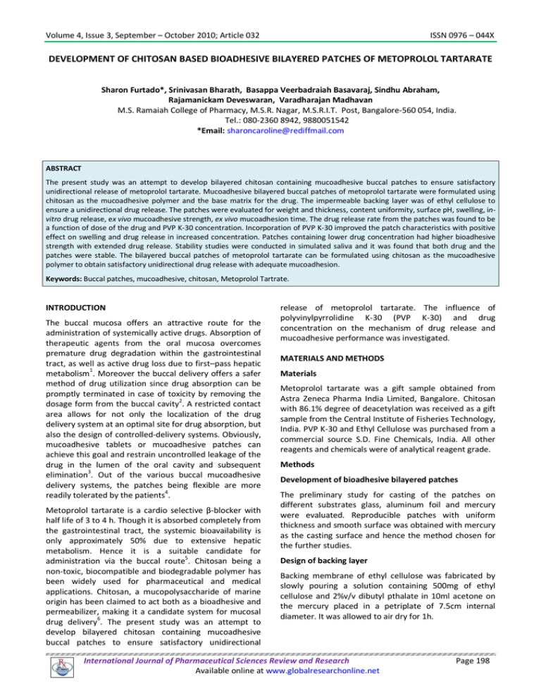
Volume 4, Issue 3, September – October 2010; Article 032 ISSN 0976 – 044X DEVELOPMENT OF CHITOSAN BASED BIOADHESIVE BILAYERED PATCHES OF METOPROLOL TARTARATE Sharon Furtado*, Srinivasan Bharath, Basappa Veerbadraiah Basavaraj, Sindhu Abraham, Rajamanickam Deveswaran, Varadharajan Madhavan M.S. Ramaiah College of Pharmacy, M.S.R. Nagar, M.S.R.I.T. Post, Bangalore-560 054, India. Tel.: 080-2360 8942, 9880051542 *Email: sharoncaroline@rediffmail.com ABSTRACT The present study was an attempt to develop bilayered chitosan containing mucoadhesive buccal patches to ensure satisfactory unidirectional release of metoprolol tartarate. Mucoadhesive bilayered buccal patches of metoprolol tartarate were formulated using chitosan as the mucoadhesive polymer and the base matrix for the drug. The impermeable backing layer was of ethyl cellulose to ensure a unidirectional drug release. The patches were evaluated for weight and thickness, content uniformity, surface pH, swelling, invitro drug release, ex vivo mucoadhesive strength, ex vivo mucoadhesion time. The drug release rate from the patches was found to be a function of dose of the drug and PVP K-30 concentration. Incorporation of PVP K-30 improved the patch characteristics with positive effect on swelling and drug release in increased concentration. Patches containing lower drug concentration had higher bioadhesive strength with extended drug release. Stability studies were conducted in simulated saliva and it was found that both drug and the patches were stable. The bilayered buccal patches of metoprolol tartarate can be formulated using chitosan as the mucoadhesive polymer to obtain satisfactory unidirectional drug release with adequate mucoadhesion. Keywords: Buccal patches, mucoadhesive, chitosan, Metoprolol Tartrate. INTRODUCTION The buccal mucosa offers an attractive route for the administration of systemically active drugs. Absorption of therapeutic agents from the oral mucosa overcomes premature drug degradation within the gastrointestinal tract, as well as active drug loss due to first–pass hepatic metabolism1. Moreover the buccal delivery offers a safer method of drug utilization since drug absorption can be promptly terminated in case of toxicity by removing the dosage form from the buccal cavity2. A restricted contact area allows for not only the localization of the drug delivery system at an optimal site for drug absorption, but also the design of controlled-delivery systems. Obviously, mucoadhesive tablets or mucoadhesive patches can achieve this goal and restrain uncontrolled leakage of the drug in the lumen of the oral cavity and subsequent 3 elimination . Out of the various buccal mucoadhesive delivery systems, the patches being flexible are more 4 readily tolerated by the patients . Metoprolol tartarate is a cardio selective β-blocker with half life of 3 to 4 h. Though it is absorbed completely from the gastrointestinal tract, the systemic bioavailability is only approximately 50% due to extensive hepatic metabolism. Hence it is a suitable candidate for administration via the buccal route5. Chitosan being a non-toxic, biocompatible and biodegradable polymer has been widely used for pharmaceutical and medical applications. Chitosan, a mucopolysaccharide of marine origin has been claimed to act both as a bioadhesive and permeabilizer, making it a candidate system for mucosal drug delivery6. The present study was an attempt to develop bilayered chitosan containing mucoadhesive buccal patches to ensure satisfactory unidirectional release of metoprolol tartarate. The influence of polyvinylpyrrolidine K-30 (PVP K-30) and drug concentration on the mechanism of drug release and mucoadhesive performance was investigated. MATERIALS AND METHODS Materials Metoprolol tartarate was a gift sample obtained from Astra Zeneca Pharma India Limited, Bangalore. Chitosan with 86.1% degree of deacetylation was received as a gift sample from the Central Institute of Fisheries Technology, India. PVP K-30 and Ethyl Cellulose was purchased from a commercial source S.D. Fine Chemicals, India. All other reagents and chemicals were of analytical reagent grade. Methods Development of bioadhesive bilayered patches The preliminary study for casting of the patches on different substrates glass, aluminum foil and mercury were evaluated. Reproducible patches with uniform thickness and smooth surface was obtained with mercury as the casting surface and hence the method chosen for the further studies. Design of backing layer Backing membrane of ethyl cellulose was fabricated by slowly pouring a solution containing 500mg of ethyl cellulose and 2%v/v dibutyl pthalate in 10ml acetone on the mercury placed in a petriplate of 7.5cm internal diameter. It was allowed to air dry for 1h. International Journal of Pharmaceutical Sciences Review and Research Available online at www.globalresearchonline.net Page 198 Volume 4, Issue 3, September – October 2010; Article 032 Design of mucoadhesive layer containing the drug Patches containing different proportions of metoprolol tartarate, chitosan and PVP K-30 were prepared by the mercury solvent casting method (Table I). One g of chitosan was dissolved in 100ml of 1.5%v/v acetic acid and sonicated until a clear solution was obtained. To improve patch performance and release characteristics, a water soluble hydrophilic additive PVP K-30, was added in different concentrations. The drug and PVP K-30 were added into the chitosan solution under constant stirring. Propylene glycol ISSN 0976 – 044X (5% v/v) was added into the solution as plasticizer under constant stirring. The viscous solution was again sonicated to homogenize and degas the solution. 10 ml of the solution was poured over the preformed backing membrane of ethyl cellulose ad allowed to dry undisturbed for 4hrs at 60°C in the oven. The dried bilayered patch was cut into dimension of 4cm2, such that each patches of F1 & F2 contained 25mg, F3 & F4 contained 50mg of the drug respectively. Table I: Composition of chitosan buccal patches of metoprolol tartarate. Batch code Ingredients F1 F2 F3 F4 Metoprolol tartarate (in mg) 25 25 50 50 Chitosan (in 1.5%v/v acetic acid) 1% 1% 1% 1% Propylene glycol (%v/v) 5% 5% 5% 5% PVP K-30 (%w/v) 0.1 0.2 0.1 0.2 Table II: Parameters of chitosan buccal patches of metoprolol tartarate. a Batch code Mass (mg)a Thickness (µm) a Drug content (%)b Surface pHb Folding enduranceb Placebo 108 ± 3 206 ± 1 0 5.9 ± 0.08 284 ± 3 F1 120 ± 2 270 ± 2 99.4 ± 0.39 5.6 ± 0.18 221 ± 5 F2 125 ± 1 282 ± 3 98.6 ± 0.64 6.3 ± 0.02 216 ± 2 F3 159 ± 3 305 ± 1 98.9 ± 0.21 6.1 ± 0.05 208 ± 4 F4 160 ± 1 316 ± 2 b = Mean ± SD, n=10 = Mean ± SD, n=3 100.7 ± 0.34 5.8 ± 0.14 196 ± 3 Table III. In vitro mucoadhesive time and mucoadhesion strength of patches. Batch code Bioadhesive strength (g) Force of adhesion (N) Bioadhesion time Placebo 15.2 0.42 740 ± 8 F1 12.4 0.12 612 ± 5 F2 11.2 0.11 547 ± 9 F3 10.3 0.10 502 ± 7 9.2 0.09 436 ± 11 F4 Mean ± SD (n=3) Table IV. Stability Study of formulated Buccal patches in simulated human saliva + → No change in appearance and color when compared with the sample at zero me interval. * → Average of Three determina ons ± S.D International Journal of Pharmaceutical Sciences Review and Research Available online at www.globalresearchonline.net Page 199 Volume 4, Issue 3, September – October 2010; Article 032 ISSN 0976 – 044X Figure 1: Swelling index of buccal patches Swelling index (%) 35 30 25 F1 20 F2 15 F3 F4 10 5 0 0 1 2 3 4 Time (h) Figure 2: Comparative release profile of metoprolol from various formulations 120 Cumulative % Drug release 100 80 F1 F2 60 F3 F4 40 20 0 0 1 2 3 4 5 Time in hours Weight and thickness of the patch The assessment of weight and patch thickness was done on 10 patches. Patches were directly weighed on a digital balance (Sartorius GE212) and patch thickness was determined by optical microscopy by taking transverse sections from different points within a patch and observing under 100X magnification. The mean and standard deviation were calculated7 Content uniformity Drug content was determined by homogenizing the patch in 100ml of isotonic phosphate buffer pH 6.8. The solution was sonicated for 30min. and filtered. The drug 8 content was then determined after proper dilution at 274nm using a UV- Visible spectrophotometer (UV 160Shimadzu, Japan). The studies were carried out in triplicate and the average values were reported. Surface pH study A combined glass electrode was used for this purpose. The patches were allowed to swell by keeping them in contact with 1ml of distilled water (pH 6.6± 0.1) for 2 h. at room temperature, and the pH was noted down by bringing the electrode in contact with the surface of the 4 patch, allowing it to equilibrate for 1 minute . Folding endurance The folding endurance of the patches was determined by repeatedly folding one patch at the same place till it broke or folded up to 300 times at the same place without breaking 8. Swelling study Buccal patch was weighed (W1), placed in a 2% w/v agar gel plate and incubated at 37± 1˚C. At regular 1 h. time intervals (up to 3 h.), the patch was removed from the petri plate and excess surface water was removed 4 carefully by blotting with a tissue paper . The swollen 9 patch was then reweighed (W2) and the swelling index was calculated from the formula, % Swelling Index = (W2 - W1)/ W1 × 100 The experiment was carried out in triplicate and the average values determined. International Journal of Pharmaceutical Sciences Review and Research Available online at www.globalresearchonline.net Page 200 Volume 4, Issue 3, September – October 2010; Article 032 In- vitro drug release studies USP apparatus type II (USP TDT 06 PL, Electrolab, Mumbai) was used to study the release of metoprolol tartarate from the patch formulations under sink conditions at 37 ± 1˚C and 50 rpm. The dissolution medium consisted of 200ml of isotonic phosphate buffer pH 6.8. A patch was applied on a glass slide in such a way that mucoadhesive layer of the patch was in contact with dissolution media and nonadhesive backing layer was fixed onto the slide with the help of cyanoacrylate adhesive. Samples (2ml) were withdrawn at predetermined time intervals and replaced with fresh 7 dissolution medium . The amount of drug released was determined spectrophotometrically at 274nm after appropriate dilution. The test was performed on three patches from each formulation. Ex vivo mucoadhesive strength The tensile strength required to detach the bioadhesive from the mucosal surface was applied as a measure of the bioadhesive performance. Fresh sheep buccal mucosa was obtained from a local slaughter house and used within 2h. of slaughter. The mucosal membrane was separated by removing the underlying fat and loose tissues. The membrane was washed with distilled water and then with isotonic phosphate buffer pH 6.8 at 37°C. Bioadhesive strength was measured on a modified physical balance. The device was mainly composed of a two arm balance. The left arm of the balance was replaced by a small stainless steel lamina vertically suspended through a wire. At the same side, a movable platform was maintained at the bottom in order to fix the model mucosal membrane. For determination of bioadhesive force, the backing membrane of the mucoadhesive patch was fixed to the stainless steel lamina using cyanoacrylate adhesive. A piece of buccal mucosa was glued to the platform. The exposed patch surface (mucoadhesive layer containing the drug) was moistened with 50µl of isotonic phosphate buffer and left for 30 s for initial hydration and swelling. The platform was then raised upward until the hydrated patch was brought into contact with the mucosal surface. A preload of 20g was placed over the stainless steel lamina for 3 min. as the initial pressure. On the right pan a constant weight of 2 g was added at 2 min. intervals. The total weight for complete detachment of the patch was recorded and the bioadhesion per unit area of the patch was calculated with the equation8: F = (Ww X G) /A, where F is the bioadhesion force, Ww is the mass applied (g), 2 G is the acceleration due to gravity (cm/s ), A is the 2 surface area of the patch (cm ). The adhesion data represents a mean of three determinations. Ex vivo mucoadhesion time The ex-vivo mucoadhesion time was evaluated after application of the patches onto freshly cut sheep buccal mucosa. The mucosa was fixed in the inner side of the ISSN 0976 – 044X beaker, above 2.5cm from the bottom with cyanoacrylate adhesive. The mucoadhesive side of each patch was wetted with one drop of isotonic phosphate buffer pH 6.8 and affixed to the sheep buccal mucosa by applying a light force with a fingertip for 30 s. The beaker was filled with 500ml of isotonic phosphate buffer pH 6.8 and temperature maintained at 37 ± 1˚C. After 2 min., a 50rpm stirring rate was applied to simulate the buccal cavity environment and patch adhesion duration i.e. the time taken for the patch to detach from the mucosa was recorded as the mucoadhesion time4. The tabulated data represents a mean of 3 determinations. Stability in simulated human saliva The stability of the patches was studied in simulated saliva solution (pH 6.8) which was prepared by dissolving NaCl (0.216g), KCl (0.964g), KSCN (0.189g), KH2PO4 (0.655g) and urea (0.200g) in 1 L of distilled water10. Patches were placed in separate petridishes containing 5ml of simulated saliva and kept in a temperature controlled oven at 37 ± 0.2˚C for 6 h. At regular intervals (0, 2, 4 and 6 h), the patches were examined for physical changes like color, texture and shape4. Drug content was determined by extracting the drug from the patch and diluting appropriately with phosphate buffer pH 6.8 and analyzed spectrophotometrically at 274 nm. RESULTS AND DISCUSSION In the present study, bilayered buccal patches of metoprolol tartarate were prepared using chitosan as the base matrix for the drug and ethyl cellulose as the backing layer. The impermeable backing layer maximized the unilateral drug concentration gradient and prolonged the adhesion due to the system being protected from saliva. The patches were casted on optimized mercury surface, using different ratios of drug to polymer and different amounts of PVP K-30 to improve the drug release and film forming properties of the patches. The prepared patches were smooth in appearance, uniform in thickness, mass, and drug content. The patch thickness ranged from 270 ± 0.82µm to 310 ± 0.46µm and mass ranged from 120 ± 0.18mg to 160 ± 0.42mg. The drug content in the buccal patches ranged from 98.6% to 100.7%, indicating favorable drug loading and uniformity of patches in terms of drug content. The surface pH of the patches was found to be between 5.6 to 6.3 and hence these patches should not cause any irritation in the buccal cavity (Table II). The swelling behaviour of the patches as a function of time shown in Fig. 1 indicated that the swelling index was greater in patches containing a higher amount of PVP K-30. Addition of this hydrophilic polymer increased the wettability and water absorbing property within the matrix. The swelling property of the patches was also evident during the drug release study. The patches retained their shape and form during the 3 h study kept on a 2% agar gel plate. International Journal of Pharmaceutical Sciences Review and Research Available online at www.globalresearchonline.net Page 201 Volume 4, Issue 3, September – October 2010; Article 032 ISSN 0976 – 044X The dissolution profile of the different formulations (Fig. 2) revealed that the slowest drug release obtained from formulation F1 and the fastest release with F4. It was studied that increasing the concentration of PVP K-30 in (F2 and F4) increased the release of the drug which could be explained by the ability of the hydrophilic polymer to absorb water, thereby promoting enhanced drug release behaviour. Moreover the polymer itself would dissolve creating more pores and channels in the matrix for the drug to leach out. It was also proved that the patches containing more drug content with 50mg. metabolism. The dose of the drug and its side effects can also be decreased with better patient compliance. (F3 & F4) showed increased and faster drug release than the patches containing 25mg of the drug (F1 & F2). This can be attributed to the water soluble nature of the drug. The drug freely dissolves in the hydrated polymer matrix and greater will be concentration of the drug in the swollen diffusional layer. Hence the concentration gradient, which is the driving force for the drug release is high leading to faster rate of drug release from these patches. The mucoadhesive time and the mucoadhesive strength of the patches were studied taking freshly obtained sheep buccal mucosa as the model mucosal membrane. The results of the study are tabulated in Table III. Increasing the drug concentration in the patch decreased both the mucoadhesive strength as well the mucoadhesive time of the patches. Incorporation of the hydrophilic polymer PVP K-30 also had a negative effect on the mucoadhesive strength and subsequently the mucoadhesive time. F1 which contained the lowest amount of the water soluble drug and hydrophilic polymer PVP K-30, showed the highest mucoadhesive strength and longest time of adhesion. The stability of the drug and the device conducted in the simulated saliva media showed no change in the physical appearance characteristics such as texture, color and shape with negligible change in the thickness when compared to the patch at zero time intervals (Table IV). The drug recovery from all the formulated patches during the study indicated maximum drug incorporation in the device. CONCLUSION From the present study, it can be concluded that the bilayered buccal patches of metoprolol tartarate can be formulated using chitosan as the mucoadhesive polymer to obtain satisfactory unidirectional drug release with adequate mucoadhesion. Development of bioadhesive buccal formulation for metoprolol tartarate may be a promising one to bypass the extensive hepatic first pass Acknowledgement: The authors are thankful to Gokula Education Foundation for providing necessary facilities to carry out the research work. REFERENCES 1. Mathiowitz E, Chickering DE, Lehr CM, Bioadhesive drug delivery systems: Fundamentals, Novel approaches and development, Marcel Dekker, Inc, 1998,563-599. 2. Choy FW, Kah HY, Kok KP, Formulation and evaluation of controlled release Eudragit buccal patches, International Journal of Pharmaceutics, 1999, 178:11–22. 3. Michael JR, Jonathan H, Michael SR, Modified release drug delivery technology, Marcel Dekker. Inc. Newyork, Basel, 2002, 354. 4. Vishnu MP, Bhupender GP, Madhabhai MP, Design and characterization f chitosan containing mucoadhesive buccal patches of propranolol hydrochloride, Acta Pharm, 2007, 57, 61-72. 5. Martindale The extra pharmacopoeia, 13th Ed, The pharmaceutical press, London. 1993, 635-636. 6. Senel S, Kremer MJ, Kas S, Wertz PW, Hincal AA, Squier CA, Enhancing effect of chitosan on peptide drug delivery across buccal mucosa, Biomaterials, 2000, 21, 2067-71. 7. Supriya SS, Nilesh SS, Sagar S, Vilasrao K, Mucoadhesive bilayered patches for administration of sumatriptan succinate, AAPS Pharm. Sci. Tech, 2008,9(3),909-916. 8. Panigrahi L, Snigdha P, Ghosal SK, Design and characterization of Mucoadhesive buccal patches of Diclofenac sodium, Indian J. Pharm. Sci, 2005, 67(3), 319-326. 9. Nakhat PD, Kondawar AA, Rathi LG, Yeole PG, Development and in vitro evaluation of buccoadhesive tablets of metoprolol tartrate, Indian J. Pharm. Sci, 2008,70 (1), 121-124. 10. Libero IG, Viviana DC, Giulia G, Maria GS, Claudio T, Ada MF, Giuseppina C, Release of naltrexone on buccal mucosa: Permeation studies, histological aspects and matrix system design, Eur. J. of Pharm and Biopharmaceutics, 2007, 67, 425-433. ************** International Journal of Pharmaceutical Sciences Review and Research Available online at www.globalresearchonline.net Page 202
