S4. SPECTROPHOTOMETRIC ANALYSIS OF PARACETAMOL TABLETS PURPOSE
advertisement
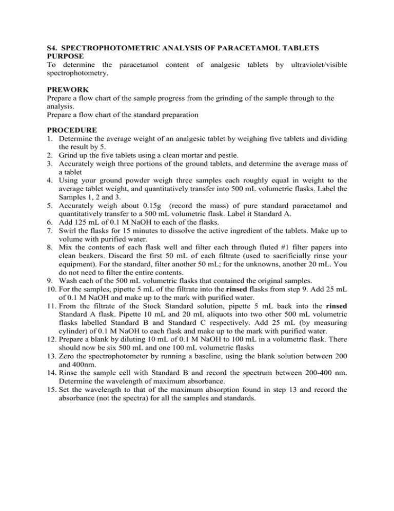
S4. SPECTROPHOTOMETRIC ANALYSIS OF PARACETAMOL TABLETS PURPOSE To determine the paracetamol content of analgesic tablets by ultraviolet/visible spectrophotometry. PREWORK Prepare a flow chart of the sample progress from the grinding of the sample through to the analysis. Prepare a flow chart of the standard preparation PROCEDURE 1. Determine the average weight of an analgesic tablet by weighing five tablets and dividing the result by 5. 2. Grind up the five tablets using a clean mortar and pestle. 3. Accurately weigh three portions of the ground tablets, and determine the average mass of a tablet 4. Using your ground powder weigh three samples each roughly equal in weight to the average tablet weight, and quantitatively transfer into 500 mL volumetric flasks. Label the Samples 1, 2 and 3. 5. Accurately weigh about 0.15g (record the mass) of pure standard paracetamol and quantitatively transfer to a 500 mL volumetric flask. Label it Standard A. 6. Add 125 mL of 0.1 M NaOH to each of the flasks. 7. Swirl the flasks for 15 minutes to dissolve the active ingredient of the tablets. Make up to volume with purified water. 8. Mix the contents of each flask well and filter each through fluted #1 filter papers into clean beakers. Discard the first 50 mL of each filtrate (used to sacrificially rinse your equipment). For the standard, filter another 50 mL; for the unknowns, another 20 mL. You do not need to filter the entire contents. 9. Wash each of the 500 mL volumetric flasks that contained the original samples. 10. For the samples, pipette 5 mL of the filtrate into the rinsed flasks from step 9. Add 25 mL of 0.1 M NaOH and make up to the mark with purified water. 11. From the filtrate of the Stock Standard solution, pipette 5 mL back into the rinsed Standard A flask. Pipette 10 mL and 20 mL aliquots into two other 500 mL volumetric flasks labelled Standard B and Standard C respectively. Add 25 mL (by measuring cylinder) of 0.1 M NaOH to each flask and make up to the mark with purified water. 12. Prepare a blank by diluting 10 mL of 0.1 M NaOH to 100 mL in a volumetric flask. There should now be six 500 mL and one 100 mL volumetric flasks 13. Zero the spectrophotometer by running a baseline, using the blank solution between 200 and 400nm. 14. Rinse the sample cell with Standard B and record the spectrum between 200-400 nm. Determine the wavelength of maximum absorbance. 15. Set the wavelength to that of the maximum absorption found in step 13 and record the absorbance (not the spectra) for all the samples and standards. CALCULATIONS 1. Calculate the concentration (in mg/L) of the original standard solution made in step 6. 2. Calculate the concentrations of the diluted standards A, B and C, (use your prework flowchart to assist). 3. Calculate the absorptivity coefficient for paracetamol, using the results from each standard 4. Plot a calibration graph of absorbance vs concentration of standards. 5. Determine the concentration of paracetamol in each of the sample solutions 6. Calculate the average paracetamol content in the tablets as %w/w, as follows (there are a number of methods to calculate the answer): • for each sample solution, apply the appropriate dilution factor to determine the original concentration of the solutions made in step 3. • for each sample solution, determine the mass of paracetamol in each flask. • to determine the %w/w, divide the mass of paracetamol in each flask by the mass of ground tablet that was used and multiply by 100. • calculate the average value and the relative precision OR • Use your flow chart to determine the amount of paracetamol in the original flask 7. Calculate the average paracetamol content in the tablets as mg/tablet by using the %w/w calculated above and the average mass of a tablet. QUESTIONS 1. Why is the blank used in this experiment not ideal? 2. Why is it necessary to prepare three or four standards for spectroscopic analyses? 3. Why was it better to carry out dilution of the standards and samples rather than simply weighing out less material initially? 4. Why does the method not just dissolve a tablet in each flask? 5. Find the structure of paracetamol. What groups in it would cause the absorption of ultraviolet radiation? 6. Find the structure of aspirin. Do you think it could be analysed in a similar manner to paracetamol? S4. RESULTS SHEET Date of analysis Identity of instruments Measurement Mass (g) 5 tablets Average tablet Sample 1 Sample 2 Sample 3 Mass of standard paracetamol Solution Blank Standard A Standard B Standard C Sample 1 Sample 2 Sample 3 Have you? Completed the instrument log Completed the sample register Completed the standard register Teachers signature Conc (mg/L) 0.00 Absorbance Date Signature
