Anti-Tropomyosin 1 (alpha) antibody ab55915 Product datasheet 3 Abreviews 4 Images
advertisement
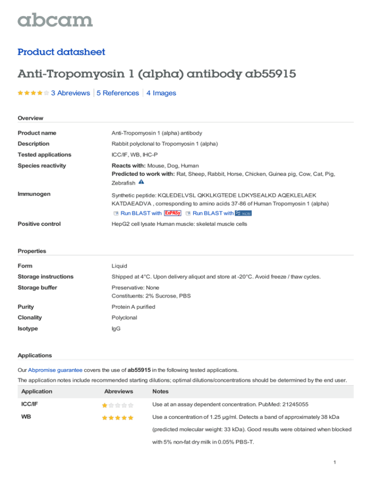
Product datasheet Anti-Tropomyosin 1 (alpha) antibody ab55915 3 Abreviews 5 References 4 Images Overview Product name Anti-Tropomyosin 1 (alpha) antibody Description Rabbit polyclonal to Tropomyosin 1 (alpha) Tested applications ICC/IF, WB, IHC-P Species reactivity Reacts with: Mouse, Dog, Human Predicted to work with: Rat, Sheep, Rabbit, Horse, Chicken, Guinea pig, Cow, Cat, Pig, Zebrafish Immunogen Synthetic peptide: KQLEDELVSL QKKLKGTEDE LDKYSEALKD AQEKLELAEK KATDAEADVA , corresponding to amino acids 37-86 of Human Tropomyosin 1 (alpha) Run BLAST with Positive control Run BLAST with HepG2 cell lysate Human muscle: skeletal muscle cells Properties Form Liquid Storage instructions Shipped at 4°C. Upon delivery aliquot and store at -20°C. Avoid freeze / thaw cycles. Storage buffer Preservative: None Constituents: 2% Sucrose, PBS Purity Protein A purified Clonality Polyclonal Isotype IgG Applications Our Abpromise guarantee covers the use of ab55915 in the following tested applications. The application notes include recommended starting dilutions; optimal dilutions/concentrations should be determined by the end user. Application Abreviews Notes ICC/IF Use at an assay dependent concentration. PubMed: 21245055 WB Use a concentration of 1.25 µg/ml. Detects a band of approximately 38 kDa (predicted molecular weight: 33 kDa). Good results were obtained when blocked with 5% non-fat dry milk in 0.05% PBS-T. 1 Application Abreviews IHC-P Notes Use a concentration of 4 - 8 µg/ml. Target Function Binds to actin filaments in muscle and non-muscle cells. Plays a central role, in association with the troponin complex, in the calcium dependent regulation of vertebrate striated muscle contraction. Smooth muscle contraction is regulated by interaction with caldesmon. In nonmuscle cells is implicated in stabilizing cytoskeleton actin filaments. Tissue specificity Detected in primary breast cancer tissues but undetectable in normal breast tissues in Sudanese patients. Isoform 1 is expressed in adult and fetal skeletal muscle and cardiac tissues, with higher expression levels in the cardiac tissues. Isoform 10 is expressed in adult and fetal cardiac tissues, but not in skeletal muscle. Involvement in disease Defects in TPM1 are the cause of cardiomyopathy familial hypertrophic type 3 (CMH3) [MIM:115196]. Familial hypertrophic cardiomyopathy is a hereditary heart disorder characterized by ventricular hypertrophy, which is usually asymmetric and often involves the interventricular septum. The symptoms include dyspnea, syncope, collapse, palpitations, and chest pain. They can be readily provoked by exercise. The disorder has inter- and intrafamilial variability ranging from benign to malignant forms with high risk of cardiac failure and sudden cardiac death. Defects in TPM1 are the cause of cardiomyopathy dilated type 1Y (CMD1Y) [MIM:611878]. Dilated cardiomyopathy is a disorder characterized by ventricular dilation and impaired systolic function, resulting in congestive heart failure and arrhythmia. Patients are at risk of premature death. Sequence similarities Belongs to the tropomyosin family. Domain The molecule is in a coiled coil structure that is formed by 2 polypeptide chains. The sequence exhibits a prominent seven-residues periodicity. Cellular localization Cytoplasm > cytoskeleton. Anti-Tropomyosin 1 (alpha) antibody images Anti-Tropomyosin 1 (alpha) antibody (ab55915) at 1.25 µg/ml + HepG2 cell lysate at 10 µg Secondary HRP conjugated anti-Rabbit IgG at 1/50000 dilution Western blot - Tropomyosin 1 (alpha) antibody (ab55915) Predicted band size : 33 kDa Observed band size : 38 kDa Gel concentration: 12% 2 ab55915 at a 1/250 dilution staining Tropomyosin 1 (alpha) in mouse heart tissue sections by Immunohistochemistry (paraffin embedded) incubated for 16 hours at +4°C. Fixed in formaldehyde, heat mediated antigen retrieval performed using citrate buffer. Blocked using 10% serum for 20 minutes at 20°C. Secondary used undiluted polyclonal Goat anti-rabbit IgG conjugated to Hilyte FluorTM 488. Immunohistochemistry (Formalin/PFA-fixed paraffin-embedded sections) - Tropomyosin 1 (alpha) antibody (ab55915) This image was kindly supplied by Dr Jinqing Li by Abreview All lanes : Anti-Tropomyosin 1 (alpha) antibody (ab55915) at 1.25 µg/ml Lane 1 : HepG2 cell lysate Lane 2 : HepG2 cell lysate with blocking peptide at 1 µg/ml Lysates/proteins at 25 µg per lane. Predicted band size : 33 kDa Western blot - Anti-Tropomyosin 1 (alpha) antibody (ab55915) Immunohistochemistry (Formalin/PFA-fixed paraffin-embedded sections) anlysis of Human Muscle lysate tissue labelling Tropomyosin 1 (alpha) with ab55915 at 8µg/ml. Positively labelled skeletal muscle cells are indicated with arrows. Magnification: 400X Immunohistochemistry (Formalin/PFA-fixed paraffin-embedded sections)-Anti-Tropomyosin 1 (alpha) antibody(ab55915) Please note: All products are "FOR RESEARCH USE ONLY AND ARE NOT INTENDED FOR DIAGNOSTIC OR THERAPEUTIC USE" Our Abpromise to you: Quality guaranteed and expert technical support 3 Replacement or refund for products not performing as stated on the datasheet Valid for 12 months from date of delivery Response to your inquiry within 24 hours We provide support in Chinese, English, French, German, Japanese and Spanish Extensive multi-media technical resources to help you We investigate all quality concerns to ensure our products perform to the highest standards If the product does not perform as described on this datasheet, we will offer a refund or replacement. For full details of the Abpromise, please visit http://www.abcam.com/abpromise or contact our technical team. Terms and conditions Guarantee only valid for products bought direct from Abcam or one of our authorized distributors 4
![Anti-Tropomyosin 1 (alpha) antibody [EPR5158] ab109505](http://s2.studylib.net/store/data/012744242_1-76009bddbb2ab7c7b2e88269514ec20a-300x300.png)
