Anti-Hsp27 antibody ab12351 Product datasheet 2 Abreviews 2 Images
advertisement
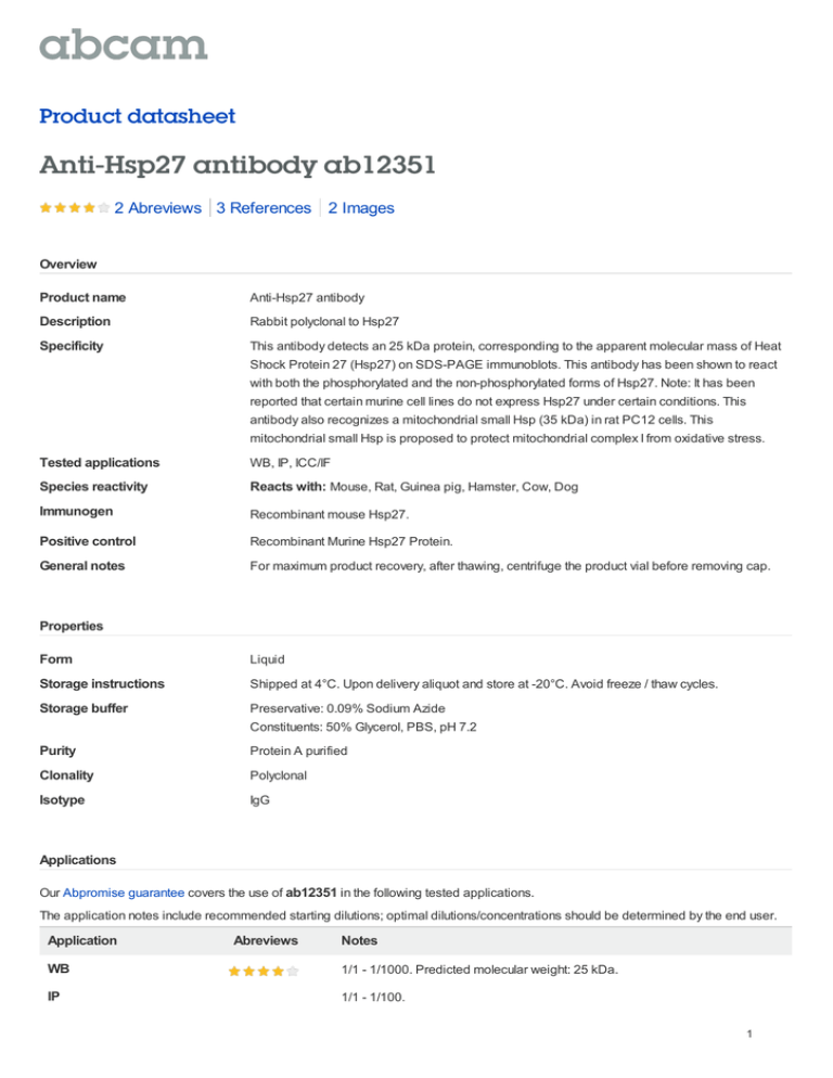
Product datasheet Anti-Hsp27 antibody ab12351 2 Abreviews 3 References 2 Images Overview Product name Anti-Hsp27 antibody Description Rabbit polyclonal to Hsp27 Specificity This antibody detects an 25 kDa protein, corresponding to the apparent molecular mass of Heat Shock Protein 27 (Hsp27) on SDS-PAGE immunoblots. This antibody has been shown to react with both the phosphorylated and the non-phosphorylated forms of Hsp27. Note: It has been reported that certain murine cell lines do not express Hsp27 under certain conditions. This antibody also recognizes a mitochondrial small Hsp (35 kDa) in rat PC12 cells. This mitochondrial small Hsp is proposed to protect mitochondrial complex I from oxidative stress. Tested applications WB, IP, ICC/IF Species reactivity Reacts with: Mouse, Rat, Guinea pig, Hamster, Cow, Dog Immunogen Recombinant mouse Hsp27. Positive control Recombinant Murine Hsp27 Protein. General notes For maximum product recovery, after thawing, centrifuge the product vial before removing cap. Properties Form Liquid Storage instructions Shipped at 4°C. Upon delivery aliquot and store at -20°C. Avoid freeze / thaw cycles. Storage buffer Preservative: 0.09% Sodium Azide Constituents: 50% Glycerol, PBS, pH 7.2 Purity Protein A purified Clonality Polyclonal Isotype IgG Applications Our Abpromise guarantee covers the use of ab12351 in the following tested applications. The application notes include recommended starting dilutions; optimal dilutions/concentrations should be determined by the end user. Application Abreviews Notes WB 1/1 - 1/1000. Predicted molecular weight: 25 kDa. IP 1/1 - 1/100. 1 Application ICC/IF Abreviews Notes Use at an assay dependent concentration. Target Function Involved in stress resistance and actin organization. Tissue specificity Detected in all tissues tested: skeletal muscle, heart, aorta, large intestine, small intestine, stomach, esophagus, bladder, adrenal gland, thyroid, pancreas, testis, adipose tissue, kidney, liver, spleen, cerebral cortex, blood serum and cerebrospinal fluid. Highest levels are found in the heart and in tissues composed of striated and smooth muscle. Involvement in disease Defects in HSPB1 are the cause of Charcot-Marie-Tooth disease type 2F (CMT2F) [MIM:606595]. CMT2F is a form of Charcot-Marie-Tooth disease, the most common inherited disorder of the peripheral nervous system. Charcot-Marie-Tooth disease is classified in two main groups on the basis of electrophysiologic properties and histopathology: primary peripheral demyelinating neuropathy or CMT1, and primary peripheral axonal neuropathy or CMT2. Neuropathies of the CMT2 group are characterized by signs of axonal regeneration in the absence of obvious myelin alterations, normal or slightly reduced nerve conduction velocities, and progressive distal muscle weakness and atrophy. Nerve conduction velocities are normal or slightly reduced. CMT2F onset is between 15 and 25 years with muscle weakness and atrophy usually beginning in feet and legs (peroneal distribution). Upper limb involvement occurs later. CMT2F inheritance is autosomal dominant. Defects in HSPB1 are a cause of distal hereditary motor neuronopathy type 2B (HMN2B) [MIM:608634]. Distal hereditary motor neuronopathies constitute a heterogeneous group of neuromuscular disorders caused by selective impairment of motor neurons in the anterior horn of the spinal cord, without sensory deficit in the posterior horn. The overall clinical picture consists of a classical distal muscular atrophy syndrome in the legs without clinical sensory loss. The disease starts with weakness and wasting of distal muscles of the anterior tibial and peroneal compartments of the legs. Later on, weakness and atrophy may expand to the proximal muscles of the lower limbs and/or to the distal upper limbs. Sequence similarities Belongs to the small heat shock protein (HSP20) family. Post-translational modifications Phosphorylated in MCF-7 cells on exposure to protein kinase C activators and heat shock. Cellular localization Cytoplasm. Nucleus. Cytoplasm > cytoskeleton > spindle. Cytoplasmic in interphase cells. Colocalizes with mitotic spindles in mitotic cells. Translocates to the nucleus during heat shock and resides in sub-nuclear structures known as SC35 speckles or nuclear splicing speckles. Anti-Hsp27 antibody images 2 Anti-Hsp27 antibody (ab12351) at 1/500 dilution + Rat keratinocytes, whole cell lysate at 10 µg Secondary HRP conjugated goat anti-rabbit antibody developed using the ECL technique Performed under reducing conditions. Predicted band size : 25 kDa Western blot - Hsp27 antibody (ab12351) Observed band size : 25 kDa Additional bands at : 120 kDa (possible non-specific binding),215 kDa (possible nonspecific binding). Exposure time : 5 minutes This image is courtesy of an anonymous Abreview All lanes : Anti-Hsp27 antibody (ab12351) at 1/1000 dilution Lane 1 : Molecular weight marker Lane 2 : Cell lysates prepared from untreated PC-12 cells Lane 3 : Cell lysates prepared from heat shock treated PC-12 cells Western blot - Hsp27 antibody (ab12351) Predicted band size : 25 kDa Please note: All products are "FOR RESEARCH USE ONLY AND ARE NOT INTENDED FOR DIAGNOSTIC OR THERAPEUTIC USE" Our Abpromise to you: Quality guaranteed and expert technical support Replacement or refund for products not performing as stated on the datasheet Valid for 12 months from date of delivery Response to your inquiry within 24 hours We provide support in Chinese, English, French, German, Japanese and Spanish Extensive multi-media technical resources to help you We investigate all quality concerns to ensure our products perform to the highest standards If the product does not perform as described on this datasheet, we will offer a refund or replacement. For full details of the Abpromise, please visit http://www.abcam.com/abpromise or contact our technical team. 3 Terms and conditions Guarantee only valid for products bought direct from Abcam or one of our authorized distributors 4
