Oral- Versus Systemic Immune Responeses Specific for
advertisement
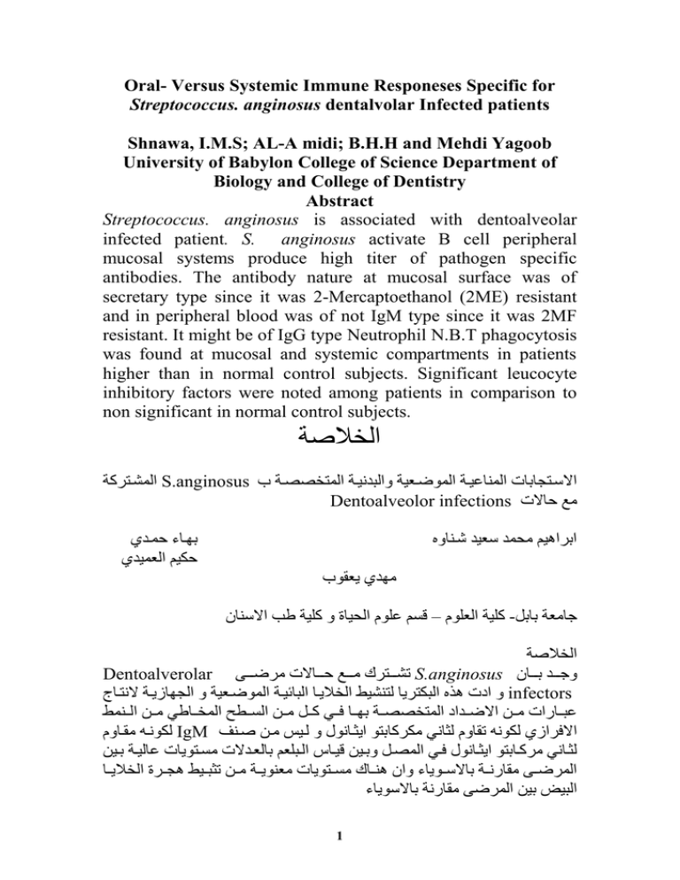
Oral- Versus Systemic Immune Responeses Specific for Streptococcus. anginosus dentalvolar Infected patients Shnawa, I.M.S; AL-A midi; B.H.H and Mehdi Yagoob University of Babylon College of Science Department of Biology and College of Dentistry Abstract Streptococcus. anginosus is associated with dentoalveolar infected patient. S. anginosus activate B cell peripheral mucosal systems produce high titer of pathogen specific antibodies. The antibody nature at mucosal surface was of secretary type since it was 2-Mercaptoethanol (2ME) resistant and in peripheral blood was of not IgM type since it was 2MF resistant. It might be of IgG type Neutrophil N.B.T phagocytosis was found at mucosal and systemic compartments in patients higher than in normal control subjects. Significant leucocyte inhibitory factors were noted among patients in comparison to non significant in normal control subjects. الخالصة االنشتجكةةاS.anginosus االستجاباب االنابيةتةاالنعيت ةةابالية ةتةاالنجخببتةا ااDentoalveolor infections معاحبال ا ا ااكاهةمامحنةاس ةةاشتابب ااااااااااااااااااااااااااااااااااااااااااااااااااااااتبحاحنتة احكةماال نةة اا ااااااااااااااااااااااااااااااااااااااااماة اي قع اا ا اةلةةاال لعما–اقسمايلعماالحةبةاباةلةةاطبااالسابناا-جبم ةاابال ا الخالصةاا Dentoalverolar اتشتتتتجك امتتتتعاحتتتتبال امكيتتتت اS.anginosusبجتتتتةااتتتتبنااا ابااد اهذ االيكجكيبالجاشةطاالخاليتبااليبيةتةاالنعيت ةةاباالااب يتةاال جتب اinfectors ييتتبتا امتتضاااليتتةاداالنجخببتتةاااتتبا تت اةتتلامتتضاالستتلماالنختتبط امتتضاالتتانطا الكع تهامقتببماIgM اال كا الكع هاتقببمالثب امككةباجعاايثتب ع ابالتةمامتضاصتا ا لثتب امكةتتباجعاايثتتب ع ا ت االنبتلاباتتةضاقةتتبعاالتتيل ماابل تةال امستتجعيب ايبلةتتةااتتةضا النكي ت امقبت تتةاابالستتعيبحاباناهاتتب امستتجعيب ام اعيتتةامتتضاتثيتتةطاهاتتكةاالخاليتتبا اليةضااةضاالنكي امقبت ةاابالسعيبحاا 1 ا Introduction The stomium is a reservoir for a variety of endogenous bacterial antigens and the port of entry for several exogenous bacterial antigens into the alimentary and respiratory systems. In normal state these antigens do not cause disease and swallowed away with saliva into the distal part of the gut (1) The protection of the stomium against bacterial invasion can be confired by either one or more of the following mechanisms mucosal barrier, continual desquamation of oral epithelium secretory IgA as well as the mucus membrane action. When an imbalance do happened between oral bacteria and immune cell cells in mucosal and systemic compartments such imbalance will lead to an immune tissue injuries or to direct tissue damage induce by oral bacteria or their host specific responses mediated by local and systemic immunity (1,2). Among the known oral bacterial pathogens are streptococci which cause several dental disease conditions (2-8). S . anginosus was reported in peridontitis patients (9). In the present work S. anginosus is being reported in cases of peridontitis, gingivitis and chronic pulpitis together with the investigation of the immune status of the patients Materials and methods 1- Patients and controls; Twelve patients out of 52 were the attendance of the surgical dental clinic, college of dentistry university of Babylon during the period Juneuary 2008 to Aprial 2009. They were diagnosed as peridontits (five cases), gingivitis (four cases) and chronic pulpitis (three cases) (2). Ten apparently healthy subjects were elected as controls) 2- Bacteriology; the dentvalveolar materials of 15 patients and 10 controls were swabbed by sterile cotton swabs into which three mls of sterile normal saline was added. Through mixing was done for these samples. Loopful inocula were taken from the swab saline mixtures and quadrate streaked onto blood agar and nutrient agar plates 2 then incubated under aerobic conditions. Biochemical identification of the pure isolates was made as in (10) 3- Immunoglobulin separation. The through mixed swabsaline mixtures became slightly opaque due to protein and cellular content (item two). These suspensions were centrifuged at 3000 rpm for five min. supernates were aspirated into sterile clean plastic tubes in three mls amounts. To these, three mls amounts of Polyethylen-glycol PEG6000% solutions was added and left at 4 C for 1 hr. The protein precipitation was settled at the bottoms of the tubes(11). Then the tubes were centrifuged at 5000 rpm for 15 min., discard supernates and dissolve precipitates in 0.5% formal saline (11,12) 4: Mucosal leucocyte separations ; The deposite of the primary swab- saline suspension (item two) was washed with saline and resuspended in three mls of saline. To this washed cell suspension three mls of 3% dextrane was added the mixture was left at room temp= 25c for 20 mins. Dextrane-lecocyte upper layer was aspirated ,tubbed into plane tube and centrifuged at 300 rpm for 15 mins.The supernate was discarded and pellet saved then resuspended, washed twice with saline and reconstituted to their original volumes in sterile saline (13) 5-Blood; Blood with and without anticoagulant in three mls amounts were collected from the brachial vein of 15 patients and ten normal subjects (14). 6: Immune function tests; The standard tube agglutination test was done as in (14). Nitroblue tetrazolium reduction test on mucosal and peripheral blood leucoytes was done following the method of Park et al (15). Leucocyte lnhibitory factors LIF were done on mucosal leucocyte by capillary method. More than 30% migration inhibition was considered as significant( 16). 3 Result: 1- Infections Agent : The dentoalveolar ,material primary plate culture revealed the growth of minute B haemolytic colonies onto blood agar plate in pure and heavy growth fashion pure culture was made and characterized as; Gram positive cocci in chains. Aerobic growth enhanced by 10% CO2 tension. Minute B-haemolytic colony morphotypes. They forms acid from lactose maltose but not from manitol and hydrolyse arginine. Negative for CAMP phenomena and for gelatine liquefaction. These characterics are consistant with S.anginosus. 2- Infection ; S.anginosus were diagnosed in association with peridontitis, gingivitis and chronic pulpitis. The immunologic aspects of these infections are present in the tables 1-4 3-Mucosal Immunoglobulin concentrations; The range of means of MIG contraction was 0.72- 0.74 g/L in patients and 0.22 g/L in controls. 4-Serum Globulin concentration the range of means of serum globulin concentration was 41.4-45.5 g/L and 36.22 for contro 5-Mucosal antibody:The S.anginosus specific mucosal antibody titers were44.8, 30 and 18.6 for peridontitis, gingivitis and chronic pulpitis as compared to normal subject these mucosal antibodies were resistant to 2ME 6: Serum antibody: The mean of the S.anginosus specific serum antibody titres were 448, 300 and 186.6 for peridontitic, gingivitis and chronic pulpitis as compared to 10 in normal subjects. These serum antibodies were resistant to 2ME. 7-Neutrophil phagoytosis:The mucosa Neutrophil N.B.T phayocytosis percentage of the patient were 43.2, 40.5 and 41.73 % for peridontitis , gingivitis and chronic pulpitis. 4 While for blood they were 36, 32.3 and 34.3% for gingivitis and chronic pulpitis as compared to in normal human subjects. Mucosal Nutrophil phagocytosis were mostely higher than that of the systemic 8-Leucoyte Inhibitory factor: The means of mucosal LIF were 52.2, 53, 5-5.3 for peridontitis gingivitis and chronic pulpity. While the mean values for systemic LIF were; 61.6, 56.25, and 67.3 for pendontitis, gingivitis and chronic pulpitis which indicate.significant inhibition percentages as compared to 99.56% in normal control subjects 5 Table (1) The immune status of S.anginosns associated peridontitis. Number Immune of cases paramear 1- MIGC 2- STPC 3- SGC 4- MIGT 5 2ME 2ME + 5- SIGT 2ME 2ME + 6-LIF M S 7- NBT M S Result Mean median range 0.37 0.84 0.79-0.84 75.32 73.7 72-82 45.5 44.3 43-47 Mean value 0.299 66.43 36.22 44.8 44.8 32 32 32-64 32-64 2 2 448 448 320 320 320-640 320-640 10 10 52.2 61.6 41.00 52.00 41-56 52-65 0.96 0.95 43.2 36 43 33 40-47 32-39 9.8 10.8 control MIGC=Mucosal Imunoglobulin concentration STPC=Serum total protein concentration SGC=Serum globulin concentration MIGT=Mucosal Immunoglobulin titer. SIGT=Serum lmmunoglobulin titer. LIF =Leucocytes lnhibitory Factor . M =Mucosal. S =Systemic N.B.T=Nitroblue tetrazolium reduction Test. Number Immune of cases paramear Result 6 Mean control value 12344 MIGC STPC SGC MIGT 2ME 2ME + 5- SIGT 2ME 2ME + 6-LIF M S 7- NBT M S Mean 0.74 72.7 41.7 30 30 median 0.64 69.7 39.5 8 8 8-64 8-64 300 300 80 80 53 56.23 55 68 40.25 32.3 range 0.74-0.78 69 -81 37-48 30 27 80-640 80-640 0.299 66.43 36.22 2 2 10 10 43- 58 36-68 0.96 0.95 30-45 27-38 9.8 10.8 Table 2 : The immune status of S.anginious associated Gingivitis MIGC=Mucosal Imunoglobulin concentration STPC=Serum total protein concentration SGC=Serum globulin concentration MIGT=Mucosal Immunoglobulin titer. SIGT=Serum lmmunoglobulin titer. LIF =Leucocytes lnhibitory Factor . M =Mucosal. S =Systemic N.B.T=Nitroblue tetrazolium reduction Test. Table 3: The immune status of S.anginosus associated chronic pulpitis 7 Number Immune of cases paramear 1- MIGC 2- STPC 3- SGC 4- MIGT 3 2ME 2ME + 5- SIGT 2ME 2ME + 6-LIF M S 7- NBT M S Result Mean median range 0.72 0.74 0.6-0. 8 71.2 70.6 70 -73 41.4 40.7 39-44 Mcan value 18.6 18.6 16 16 8-32 8-32 2 2 186 186 160 160 160-320 160-320 10 10 52- 57 57-66 0.96 0.95 36-43 32-37 9.8 10.8 55.3 57. 3 41.67 34.33 52 60 46 37 0.299 66.43 36.22 MIGC=Mucosal Imunoglobulin concentration STPC=Serum total protein concentration SGC=Serum globulin concentration MIGT=Mucosal Immunoglobulin titer. SIGT=Serum lmmunoglobulin titer. LIF =Leucocytes lnhibitory Factor . M =Mucosal. S =Systemic N.B.T=Nitroblue tetrazolium reduction Test. Table 4 : The immune status of S.anginosus associated dentoalar infections 8 Nof cases 12 Immune paramear MIGC STPC SGC MIGT 2ME2ME + SIGT 2ME 2ME + LIF M S NBT M S Mean of Peridontitis gngivits chronic pulpitis 0.73 0.74 0.72 75.32 72.7 71.2 45.4 41.7 41.4 Mcan value 0.299 66.43 36.22 44.8 44.8 30 30 18.6 18.6 2 2 448 448 300 300 186.6 186.6 10 10 52.2 61. 6 53 56.25 55.3 67.3 0.96 0.95 43.2 36 40.25 92.3 71.7 34.3 9.8 10.8 MIGC=Mucosal Imunoglobulin concentration STPC=Serum total protein concentration SGC=Serum globulin concentration MIGT=Mucosal Immunoglobulin titer. SIGT=Serum lmmunoglobulin titer. LIF =Leucocytes lnhibitory Factor . M =Mucosal. S =Systemic N.B.T=Nitroblue tetrazolium reduction Test 9 control Discussion S. anginosus is being reported in association with peridontitis gingivitis and chronic pulpitis (9,17,18,19)the rise in S.anginiosus specific antibody titer may indicate B cell poly clonal activiting epitope and /or Th 2 cell polyclonal activitating epitope(20). In addition to an epitope activating Toll-like receptor that reacts with pattern recognition structure on S anginousus as indicated by the increase in NBT in patient (21,22).As well as LIF inducing T Cell epitope(21,22) as revealed by significant migration inhibition of lecocytes in patient groups. The 2ME resistance in mucosal antibodies means that they are of secretary type (23) While 2ME resistance in serum antibodies indicate that these antibodies were not of IgM type (22)thus, on summing up the basic immune features of the dentoalveolar infections with S.anginosus, one may state that 1. S. anginosus is assotiated causal with dentoalveolar infection 2. it induces an increase in mucosal globulin concentrations to levels higher than those of normal control subjects 3. it activate neutrophil phogoytosis 4. it stimulate B cell, th1 and th2 cell responses 5. it stimulate LIF cytokine production in significant level both in mucosal and peripheral blood compartments . References 1- Parslow, T.G.; Stites,D.P.; Terr A.I and Imboden J.B. 2001.Medical Immunology Alange Medical books N.Y. 2- Samaranayake, L.P. and Jones, B.M. 2002. Essential Microbcology for Dentistry Churchill- Livingstone, London , 224- 231. 3- Cicek , Y. and Ozgoz, M.J Contemp. Dent Pract 2004, 5(3): 1-5 4- Porta, G.; Rodriguec- Carballeira; Gomec, Salavert, M. and others. Eur. Respire. J. 1998. 12:357-362. 10 5- Iopez, D.F.P.Rev. Soc. Bol. Ped.2005, 11 (3): 153-7 6- Znu,H.;Wilcox, M.D.p. and Knox, K.W. Int.J. Syst. Evol. Microbiol. 2000. 50 : 55-61 7- Rigante , D.; Spanu, T; Nanni, L.; Tornesello, A. and others. BMC Infections Disease 2006, 6: 61- 64. 8- Paster, B.J; Olson, I.; AAS, J.A. and Dewhirst; F.E. peridontol 2006,42: 80-87. 9- Sungano, W.; Yokoyama, k.; Oshikawa, M., Kumagai, k; Takane, M. and others. J. Orl. Sci 2003, 45(4)181-184. 10MaCfaddin,J.F.2000. Biochemical Identification of Bacteria 2nd .ed. Williams and Wilkins company Co. 11Johnstone , A, and Thorpe, R. 1982. Immunochemistry In Practice. Blackwell scientific publications, London ., 64- 70 12-Shnawa , I.M.S; and AL-Sadi,M.A.K:Gut Mucosal Lmmunoglobulin Separation ,Partial characterization .Irq. J.Microbiol.13 (3) :57- 70. 13-Metacalf, J.A.; Gallin J.I.; Nausect W.M and Root, R.K. 1986. laboratory Manual of Neutrophil Fanction. Raven Press, N-Y., 2-3 14-Garvey, S.J.; Cremer N.E. and Sussdoof D.H. 1977. Methods In Immunology 3rd W.A. Benjamin, NC, London 15-Park, B.H; Fikrina , S.M and Wick, S.E.J. Lancet. 1968, 2:532 – 534. 16-Soberg, M.Act.Med- Scand 1968. 184:235 17-Ruoff K.L. Clin Microbil Rev. 1988, 1(1): 102-108 18-Brook;G.F; Butel J.S. and Morse S.A. 2007. Jawets, Melnde and Adelbergs Medical Microbiology 24th ed. Megrar. Hill Co. N.Y. 19-Rozkiewiz, D.; Daniluk, T; Sciepuk, M.; Zaaremba, ML., Cylwik. Akicka, D., and others Adv. Med. Sci. 2006 s1 a 20-Zubler, R.H. 1998 ;Antigens, In Delves, J,. and Roitt, I. I . Encyclopedia of Immunology . 2nd ed . Academic Press, London , 214 – 218. 21--Delves.P.J., Martin, S.J.; Burton D.R. and Roitt I.M. 2006 Roitts Essertial Immunology 11th ed Blackwell Scien tifie Publishing . 11 22-Male, D; Brostoff, I.; Roth, D.B and Roitt I. 2006 Immunlogy 7th ed Mosby. Elsevier Canade. 23-Tomasi T.B 1976. the Immune System of Secretions. Prentice- Hall Inc W.J. 12
