Isolation of Anaerobic Microbiota of Slug Intestinal Tract
advertisement
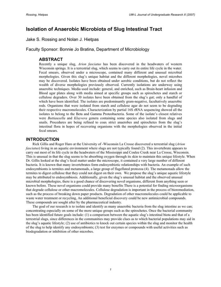
Rossing, Hietpas UW-L Journal of Undergraduate Research X (2007) Isolation of Anaerobic Microbiota of Slug Intestinal Tract Jake S. Rossing and Nolan J. Hietpas Faculty Sponsor: Bonnie Jo Bratina, Department of Microbiology ABSTRACT Recently a unique slug, Arion fasciatus has been discovered in the headwaters of western Wisconsin springs. It is a terrestrial slug, which seems to carry out its entire life cycle in the water. Fecal smears, observed under a microscope, contained many different and unusual microbial morphologies. Given this slug’s unique habitat and the different morphologies, novel microbes may be discovered. Isolates have been obtained under aerobic conditions, but do not reflect the wealth of diverse morphologies previously observed. Currently isolations are underway using anaerobic techniques. Media used include: general, and enriched, such as Brain-heart infusion and Blood agar plates along with media aimed at specific groups such as spirochetes and starch or cellulose degraders. Over 30 isolates have been obtained from the slug’s gut; only a handful of which have been identified. The isolates are predominantly gram-negative, facultatively anaerobic rods. Organisms that were isolated from starch and cellulose agar do not seem to be degrading their respective macromolecules. Characterization by partial 16S rRNA sequencing showed all the isolates to belong to the Beta and Gamma Proteobacteria. Some of the isolate’s closest relatives were Buttiauxella and Kluyvera genera containing some species also isolated from slugs and snails. Procedures are being refined to coax strict anaerobes and spirochetes from the slug’s intestinal flora in hopes of recovering organisms with the morphologies observed in the initial fecal smears. INTRODUCTION Rick Gillis and Roger Haro at the University of -Wisconsin La Crosse discovered a terrestrial slug (Arion fasciatus) living in an aquatic environment where slugs are not typically found (2). This invertebrate appears to carry out most of its life cycle in the headwaters of the Mississippi and Coulee Creek near La Crosse, Wisconsin. This is unusual in that the slug seems to be absorbing oxygen through its skin to maintain this unique lifestyle. When Dr. Gillis looked at the slug’s fecal matter under the microscope, it contained a very large number of different bacteria. It is known that many invertebrates form endosymbiotic relationships with bacteria. An example of such endosymbionts is termites and metamonads, a large group of flagellated protozoa (4). The metamonads allow the termites to digest cellulose that they could not digest on their own. We propose the slug’s unique aquatic lifestyle may be attributed to endosymbiosis. Additionally, given the slug’s unusual habitat and the observed unusual microbial morphologies, there is a good chance of discovering novel organisms, different from anything seen or known before. These novel organisms could provide many benefits There is a potential for finding microorganisms that degrade cellulose or other macromolecules. Cellulose degradation is important in the process of bioremediation, such as the process of breaking down paper products. Degradation of other macromolecules could be applicable to waste water treatment or recycling, An additional beneficial discovery could be new antimicrobial compounds. These compounds are sought after by the pharmaceutical industry. The goal of our research is to isolate and identify as many anaerobic bacteria from the slug intestine as we can; concentrating especially on some of the more unique groups such as the spirochetes. Once the bacterial community has been identified future goals include: (1) a comparison between the aquatic slug’s intestinal biota and that of a terrestrial slugs, since differences in the communities may provide clues as to which bacterial populations may aid in the slug’s aquatic lifestyle; (2) use of antibiotics to knock out certain species within the slug and monitor the health of the slug to help identify any endosymbionts; (3) test for enzymes or compounds with useful activities such as biodegradation or inhibition of other microbes. 1 Rossing, Hietpas UW-L Journal of Undergraduate Research X (2007) MATERIALS AND METHODS Collection Slugs were collected from springs near Blue Bird Campground, located northeast of La Crosse, WI. Dissection The slugs were surface sterilized by soaking them for 3 minutes in a 0.5% solution of chlorohexadione. The slugs were then rinsed twice with sterile deionized water. In an anaerobe chamber, the entire digestive system was removed with a sterile scalpel, weighed, and added to a 9 mL dilution blank of standard methods buffer. Isolation The mixture was emulsified with a sterile mortar and pestle, and serially diluted to 10-8, and spread plated to various media. Plates were incubated until sufficient growth for colony isolation appeared (1-5 days). Isolated colonies were picked with a sterile wood applicator to duplicate plates. The first plate was grown aerobically and the second was grown anaerobically. Media Media used for isolation: glucose yeast peptone tryptone (GYPT), mineral salts plus cellulose, starch agar, bile esculin, brain heart infusion (BHI), mineral salts plus cellulose and yeast extract (MSYEC), cooked meat broth, blood agar, pectin casein nitrate (PCN), Barker’s methanogen enrichment media, NOS spirochete media and veal infusion. Polymerase Chain Reaction (PCR) and Sequence Analysis Isolated colonies were lysed for PCR using either Lyse-N-Go (Pierce, Rockford, IL) or distilled water at 95oC for 5 minutes. The lysate was added directly to the PCR reaction along with primers (8F and 1492R) (1). Amplification products were confirmed by running agarose gel electrophoresis. The amplification products were then sequenced for identification using the University of Wisconsin – Madison Biotechnology Center. Sequences were analyzed using the Ribosomal Database Project (RDP) Sequence Match program (5). RESULTS & DISCUSSION Using an anaerobe chamber, we were able to isolate different strains of bacteria while using the same media as was used in previous aerobic isolations (Lamberson, pers. comm.). Most of the isolates obtained were gram negative, facultatively anaerobic rods (Table 1). Phylogenetic analysis showed some of the isolates found were related to Buttiauxella and Kluyvera, genera that have been previously isolated from mollusks (Table 2). Buttiauxella and Kluyvera are enteric bacteria whose general habitat is in the intestines of organisms including slugs (3). This makes them likely candidates for endosymbionts and so will be followed up in future studies. Thus far our endeavors to isolate bacteria with the unusual morphologies have been unsuccessful. Phylogenetic analysis of some of the isolates also indicated that these were not novel organisms (all were greater than 90% similarity to 16S ribosomal RNA from cultured organisms) We, therefore, intend to continue the isolations using new media suggested by phylogenetic studies (Lamberson, per. comm.) to enrich for and culture novel organisms. 2 Rossing, Hietpas UW-L Journal of Undergraduate Research X (2007) Table 1. Characterization of anaerobic isolates from Arion fasciatus Isolate B1-4(1) B1-4(2) B1-5(1) B1-5(2) B1-5(3) B1-6(1) B1-8(1) V1-6(1) V1-6(2) V1-6(3) V1-8(1) V1-8(2) MSYEC1-7(1) MSYEC1-7(2) MSYEC1-7(3) Sven B B2-2(1) B2-2(6) B2-2(9) B2-2(10) B2-4(1) B2-4(3) B2-4(4) B2-4(5) B2-4(10) PCN (L) PCN (B) Spyro A1 A2 A3 Media BHI BHI BHI BHI BHI BHI BHI Veal Infusion Veal Infusion Veal Infusion Veal Infusion Veal Infusion MSYEC MSYEC MSYEC GYPT BHI w/Km BHI w/Km BHI w/Km BHI w/Km BHI w/Km BHI w/Km BHI w/Km BHI w/Km BHI w/Km PCN PCN NOS Spjrochete media Blood Agar w/Km Blood Agar w/Km Blood Agar w/Km Gram stain nd nd nd nd nd nd nd nd nd nd nd nd nd nd nd Gr (-) Gr (-) Gr (-) Gr (-) Gr (-) Gr (-) Gr (-) Gr (-) Gr (-) Gr (-) Gr (-) Gr (-) Morphology nd nd nd nd nd nd nd nd nd nd nd nd nd nd nd Rod Rod Rod Rod Rod Rod Rod Rod Rod Rod nd nd Oxidase nd nd nd nd nd nd nd nd nd nd nd nd nd nd nd nd Neg Neg Neg Neg Neg Neg Neg Neg Neg Pos Neg Catalase nd nd nd nd nd nd nd nd nd nd nd nd nd nd nd nd Pos Pos Pos Pos Neg Neg Neg Neg Neg Neg Neg Gr (-) nd nd nd nd nd nd nd nd nd nd nd nd nd nd nd 3 Rossing, Hietpas UW-L Journal of Undergraduate Research X (2007) Table 2. Phylogenetic analysis to characterize selected isolates from Arion fasciatus. Isolate sequences James Frau X Z Gerry A1 A3 B2-2(2) B2-2(6) B2-2(9) B2-2(10) PCN (L) PCN (B) B2-4(4) B2-4(1) a Closest relative Uncultured org. rRNA334 Delftia sp LFJ11- 1 Buttiauxella spp.a Buttiauxella sp. B22a Comanonas sp. iCTE630 Citrobacter freundii Kluyvera cryocerscens Aeromonas sobria Aeromonas hydrophila Aeromonas salmonicida Aeromonas media Pseudomonas tolaasii Stenotrophomonas maltophilia Aeromonas encheleia Aeromonas popoffii % Similarity 98.3 95.3 94.2 94.0 90.9 98.5 97.8 99.6 97.2 100.0 99.3 99.2 99.5 100.0 100.0 The genera Buttiauxella and Kluyvera is known to contain species the were isolated from other gastropods including slugs. ACKNOWLEDGEMENTS This research was funded in part by a Fall 2006 UW - La Crosse Undergraduate Research Grant. Travel expenses were paid in part by UW-La Crosse College of Science and Health. Thanks to Mallory “Princess” Lamberson and Jim Parejko. Thank you Rick Gillis and Roger Haro. REFERENCES 1. 2. 3. 4. 5. Eden, P.A., T.M. Schmidt, R.P. Blakemore, and N.R. Pace. 1991. Phylogenetic analysis of Aquaspirillum magnetotacticum using polymerase chain reaction amplified 16S rRNA-specific DNA. Int. J. Sys. Bacteriol. 41:324-325. Haro, R. J., R. Gillis and S.T. Cooper. 2004. First report of a terrestrial slug (Arion fasciatus) living in an aquatic habitat. Malacologia 45:451-452. Janda, J. M. New members of the family Enterobacteriaceae. In M. Dworkin, S. Falkow, E. Rosenberg, KH. Schleifer and E. Stackebrandt, (ed.), The prokaryotes. Springer New York, New York. Madigan, Michael T., J.M. Martinko, and J. Parker. 1997. Biology of microorganisms. Eighth edition. p. 352. Prentice Hall, Upper Saddle River, NJ. Maidak, B.L., G. J. Olsen, N. Larson, R. Overbeek, M. J. McCaughey, and C.R. Woese. 1996. The Ribosomal Database Project (RDP). NA Res. 24:82-85. 4
