Perovskite to Postperovskite Transition in NaFeF
advertisement
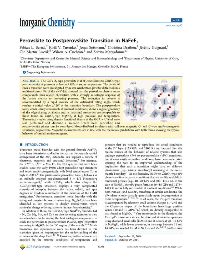
Article pubs.acs.org/IC Perovskite to Postperovskite Transition in NaFeF3 Fabian L. Bernal,† Kirill V. Yusenko,† Jonas Sottmann,† Christina Drathen,‡ Jérémy Guignard,‡ Ole Martin Løvvik,§ Wilson A. Crichton,‡ and Serena Margadonna*,† † Chemistry Department and Centre for Material Science and Nanotechnology and §Department of Physics, University of Oslo, NO-0315 Oslo, Norway ‡ ESRFThe European Synchrotron, 71, Avenue des Martyrs, Grenoble 38043, France S Supporting Information * ABSTRACT: The GdFeO3-type perovskite NaFeF3 transforms to CaIrO3-type postperovskite at pressures as low as 9 GPa at room temperature. The details of such a transition were investigated by in situ synchrotron powder diffraction in a multianvil press. Fit of the p−V data showed that the perovskite phase is more compressible than related chemistries with a strongly anisotropic response of the lattice metrics to increasing pressure. The reduction in volume is accommodated by a rapid increase of the octahedral tilting angle, which reaches a critical value of 26° at the transition boundary. The postperovskite form, which is fully recoverable at ambient conditions, shows a regular geometry of the edge-sharing octahedra and its structural properties are comparable to those found in CaIrO3-type MgSiO3 at high pressure and temperature. Theoretical studies using density functional theory at the GGA + U level were also performed and describe a scenario where both perovskite and postperovskite phases can be considered Mott−Hubbard insulators with collinear magnetic G- and C-type antiferromagnetic structures, respectively. Magnetic measurements are in line with the theoretical predictions with both forms showing the typical behavior of canted antiferromagnets. ■ INTRODUCTION Transition metal fluorides with the general formula AM2+F3 have been intensively studied in the past as the versatile spatial arrangement of the MF6 octahedra can support a variety of electronic, magnetic, and structural behaviors.1 For instance, the KM2+F3 (M2+ = Mn, Fe, Co, Ni) systems that have been studied since the early 1960s adopt perovskite-type structures and order antiferromagnetically with Néel temperatures TN as high as 280 K.2 The pseudocubic perovskite KCuF3 behaves as an orbitally ordered one-dimensional S = 1/2 Heisenberg antiferromagnet,3 while KCrF3, which also adopts the KCuF3(tI20)-type structure, displays a very complicated scenario of interplay between the lattice, orbital, and spin degrees of freedom reminiscent of the behavior of the CMR manganites.4 Furthermore, transition metal fluorides with the tetragonal tungsten bronze structure (e.g., K0.6FeF3) have been identified as key systems to display multiferroism where particular charge ordering patterns induce ferroelectricity.5 In addition to these, the GdFeO3-type NaM2+F3 fluorides (M = Ni, Co, Mg, Mn, and Zn) are also receiving attention as they are considered to be among the best analogous compounds to study the perovskite to postperovskite (CaIrO3-type) transition occurring in MgSiO3 in the D″ region of the mantle.6−9 Much theoretical and experimental work has been devoted to this transition given its importance for the understanding of the structure of the deep Earth.10−12 However, further advances are impeded by the extreme conditions of temperature and © 2014 American Chemical Society pressure that are needed to reproduce the actual conditions at the D″ layer (125 GPa and 2500 K) and beyond. For this reason studies of the behavior of related systems that also undergo perovskite (Pv) to postperovskite (pPv) transitions, but at more easily accessible conditions, have been undertaken opening the way to an improved understanding of the implication that such a transition might have on different phenomena (e.g., seismic anisotropy) occurring at the core− mantle boundary.12 In the fluorides, the Pv to CaIrO3-type pPv phase transition occurs at conditions that are readily available to multianvil presses (e.g., 10−20 GPa and 800−1473 K). In the case of NaNiF3, the pPv phase forms at 16−18 GPa and 1273− 1473 K and is fully recoverable at ambient conditions.8,9 While both NaCoF3 and NaZnF3 transform at similar conditions, the pPv phase is only partially quenchable upon decompression at room temperature.6−9,12−15 In all cases, the Pv−pPv transition is accompanied by relatively small volume changes (1−2%) and the Clapeyron slopes of the boundaries have large positive values (10 and 17 MPa/°C) which are directly comparable to that found in MgSiO3.14 Very importantly, in the fluorides, the Pv to pPv transition can also be observed at room temperature using diamond anvil cells (DACs) and it occurs at 28−30 GPa in NaMgF3, while lower pressures, in the range between 11 and 14 GPa, are needed for M = Ni, Co, and Zn.8,9,13 Further laser Received: September 12, 2014 Published: October 29, 2014 12205 dx.doi.org/10.1021/ic502224y | Inorg. Chem. 2014, 53, 12205−12214 Inorganic Chemistry Article collected during all the steps of the synthesis and were recorded using a linear pixelated GOS detector from Detection Technology, running 32 sequential exposures at 10 Hz at 32 s intervals and mounted to intercept the downstream diffraction from the horizontal anvil gap at a distance of ∼1180 mm from the sample. The detector, beam-normal plane was mechanically corrected for tilt and rotation and the detector position and zero offset was calibrated against SRM660a, at a monochromatic wavelength of 0.3757 or 0.2254 Å, selected from the emission of a U18 undulator (6.1−6.2 mm gap) by a Cinel Si(111) double-crystal monochromator. Selected data for refinement were also collected using a Tl:NaI scintillator counter, with a 0.2 mm pinhole and receiving slits at intermediate distance, which acted as collimators of background scattering coming from the cell assembly. In this case, the effective radius of the pseudorotation detector arm was 1350 mm. Collimated beam sizes were 0.5 mm horizontal by 1 mm, or the vertical anvil gap, when smaller. Ambient pressure high-resolution synchrotron X-ray diffraction experiments were performed on beamline ID31 at the ESRF. Finely ground samples of both Pv and recovered pPv NaFeF3 were sealed in 0.5 mm diameter thin-wall glass capillaries. Diffraction profiles were recorded at various temperatures in the range T = 10−300 K (λ = 0.399 92 Å). The data were binned in the 2θ range 0.1−30° to a step of 0.002°. All the powder diffraction profiles collected at ID06 and ID31 were analyzed using the GSAS suite of Rietveld programs.21,22 The dc magnetization measurements of Pv and pPv NaFeF3 were performed on a Quantum Design PPMS system and on a Quantum Design MPMS magnetometer, respectively. The temperature dependence of magnetization was determined following zero-field-cooled and field-cooled (ZFC, FC) protocols in magnetic fields H = 0.01, 0.1, and 1 T. Magnetic hysteresis loops up to H = 9 T for Pv and H = 5 T for pPv were collected at various temperatures. Electronic structure calculations were performed with the Vienna ab initio simulation package (VASP)23,24 using the PBE generalized gradient approximation (GGA).25 The cutoff energy of the plane wave basis set expansion was 500 eV, and the density of the k-points was determined by a maximum distance of 0.25 Å−1. All structures were relaxed with remaining forces lower than 0.05 eV/Å, using a quasiNewton method. heating in DACs of the so-formed pPv phases induces a transition to a yet unknown structure in NaMgF3 (a so far elusive possible post-pPv phase),7 to decomposition to CsCltype NaF + MgF2, and to more complex disproportionation in NaCoF3.8 Up to now, accurate structural refinement of pPv fluoride phases has been achieved only for NaNiF3.9 NaFeF3 is another member of the fluoroperovskite family and is attracting interest as an efficient and stable cathode material for secondary Na ion batteries. Very surprisingly, little is currently known on the structural and magnetic properties of NaFeF3 as a function of temperature and pressure. At ambient conditions, it adopts the GdFeO3-type structure which was determined by single-crystal methods by Benner and Hoppe.16 Careful inspection of the reported lattice parameters and atomic coordinates quickly identify that NaFeF3 possesses all the important prerequisites (values of tolerance factor, tilting angle, and polyhedral volume ratio) to undergo a transition to a CaIrO3-type phase at very amenable conditions. Indeed, we were recently successful in observing the Pv−pPv transition between 10 and 12 GPa in single-crystal samples of NaFeF3, which were hydrostatically compressed using DACs at room temperature.17 Very interestingly, further increase in pressure up to 20 GPa results in the stabilization of a postpostperovskite Sb2S3-type structure. This is the first experimental observation of any higher pressure forms beyond CaIrO3 type in any system and is calculated as a stable enthalpy-driven form only for Al2O3 (at 3.6 Mbar),18 while it is calculated as metastable in NaMgF3.19 The equivalent pressure, for MgSiO3, is estimated at 1.6 TPa, though this is also estimated to be metastable.20 The understanding of such a transition allows for the construction of a unified framework where possible structural sequences in perovskites and the competitive sesquioxides can be predicted beyond pPv, over a much wider range of chemistries. The realization that NaFeF3 undergoes the transformation to pPv at lower pressures (in single-crystal specimens at room temperature (RT)) than other members of the family has prompted us to further investigations with the aim of defining a general description of the Pv−pPv transition in fluoroperovskites to compare and contrast with those proposed for the ABO3 analogues. Here we report on the synthesis of polycrystalline pPv NaFeF3 using multianvil apparatus together with a detailed description of the structural and magnetic properties of both Pv and pPv forms of NaFeF3. The results of synchrotron powder X-ray diffraction measurements, magnetometry, and ab initio density functional theory (DFT) calculation will be discussed. ■ ■ RESULTS Perovskite NaFeF3: Crystal Structure and Magnetic Properties. Inspection of the high-resolution synchrotron powder diffraction profile at room temperature readily confirms the synthesis of a single-phase polycrystalline sample of NaFeF3, adopting the orthorhombic GdFeO3-type structure (a = c ∼ √2ap, b ∼ 2ap, where ap is the lattice constant of the pseudocubic perovskite cell). Analysis by the Le Bail pattern decomposition technique resulted in lattice parameters a = 5.66119(2) Å, b = 7.88006(2) Å, and c = 5.48359(4) Å (space group Pnma), in agreement with previous work.16 The final Rietveld refinement shows that the structure can be described as a three-dimensional network of almost regular corner-sharing FeF6 octahedra (Figure 1). The values of the metal−fluorine distances are consistent with the presence of the weakly Jahn−Teller distorted Fe2+ ions in high-spin (HS) electronic configuration and can be divided into medium bonds (2.072(1) Å) along the b-axis and alternating longer (2.085(1) Å) and shorter (2.052(1) Å) bonds in the ac plane, resulting in a very small value of the octahedral distortion parameter, Δd = 42.2 × 10⁻⁶. The orthorhombic distortion is then purely driven by the relative sizes of the ions as indicated by the calculated tolerance factor, t = 0.85. It is well-known that the value of t ∼ 0.9 is considered the lower boundary for the stabilization of cubic perovskites. The small size of the 8-fold coordinated Na+ cation induces tilting of the octahedra and distortion from the ideal cubic Pv EXPERIMENTAL SECTION Polycrystalline samples of Pv NaFeF3 samples were synthesized according to ref 16. Degassed NaF, Fe, and FeF3 powders were finely ground and mixed in a 3:2:1 ratio. The so-obtained mixtures were then pelletized and loaded into an Fe tube which was used as a reaction vessel. All the manipulations were carried out in an Ar-filled glovebox, given the hygroscopic nature of NaF, and to avoid exposure to oxygen and/or moisture. The Fe tube was tightly closed and then heated under a constant nitrogen flow at 750 °C for 8 days. The cubic multianvil press available at ID06LVP at the ESRF was used for the synthesis of pPv NaFeF3. A 2 mg sample of the original Pv phase was packed into an hBN (Goodfellow) capsule, before being included into the 10/4 windowed Cr:MgO assembly (Ceramic Substrates). Pressure was generated using the 2000 ton MAVO press in 6/8(×32) mode, with carbide anvils. Pressures were estimated using the equation of state of BN, and a graphite furnace was used to achieve high temperatures. Synchrotron powder diffraction data were 12206 dx.doi.org/10.1021/ic502224y | Inorg. Chem. 2014, 53, 12205−12214 Inorganic Chemistry Article takes the value of 4.2 in Pv NaFeF3. Finally, the calculated bond valence sum (BVS) values of Na and Fe are consistent with the valence states Na+ and Fe2+. The molar magnetic susceptibility χM measured as a function of temperature in an applied magnetic field H = 1 T on powder samples of Pv NaFeF3 is presented in Figure 2. The data show a sharp increase of χM below 90 K, providing a clear signature of the onset of long-range magnetic ordering. At temperatures lower than 20 K, χM starts decreasing. It is well-known that NaBF3 fluorides adopting the GdFeO3 structure (B = Ni, Mn, Co) can be described as G-type antiferromagnets.27−30 Weak ferromagnetism is observed below the Néel temperature TN, which is associated with the presence of spin canting. Our data point toward a scenario where also NaFeF3 can be described as a weak ferromagnet with TN = 90 K in analogy with the other members of the family. As expected for this type of magnetic ordering, the temperature dependence of the ZFC and FC magnetization measured at the lower magnetic fields of 0.01 and 0.1 T show divergence below 90 K (Figure 2 and Supporting Information, Figure S1). Magnetization vs H curves were collected at different temperatures. At 4 K, a small hysteresis loop with coercive field of 2.4 mT is observed and the magnetization reaches the value of 0.36 μB/Fe atom at 9 T (Figure 2). The lack of Curie−Weiss behavior at temperatures well above TN and the shape of the M vs H curve at 300 K demonstrate the absence of paramagnetic behavior, even at room temperature, and point toward the existence of an underlying magnetic contribution which could be due to the presence of impurities. Indeed, small traces (<0.5%) of unreacted α-Fe are visible in the high-resolution synchrotron diffraction profiles. Perovskite NaFeF3: Electronic Structure. To get further insight into the properties of the Pv phase of NaFeF3, electronic structure calculations were performed. They were based on density functional theory at the GGA + U level to take into account the highly correlated nature of the Fe2+ d-electrons. Four different spin-ordered structures were considered as possible ground states: (i) F-type spin ordering with intra- and interlayer ferromagnetic arrangement; (ii) A-type spin ordering with intralayer ferromagnetic and interlayer antiferromagnetic spin arrangements; (iii) C-type spin ordering in which the magnetic centers interact antiferromagnetically with the inplane nearest neighbors and ferromagnetically with those out of plane; (iv) G-type spin ordering with antiferromagnetic exchange with all the nearest neighbors (Supporting Information, Figure S4). The analysis of the exchange interactions was performed using the nearest-neighbor Heisenberg Hamiltonian which allows their evaluation from total energy calculations.31,32 Two magnetic exchange parameters were considered to describe the interlayer (Jac) and the intralayer (Jb) coupling in the orthorhombic Pv unit cell. The Hubbard parameter U was varied evenly in steps of 2 eV from 0.0 to 8.0 eV with the Hund’s exchange parameter of JH = 0.98 eV obtained from former local spin density approximation (LSDA) calculations on KFeF3.33 The calculated electronic energies of the different magnetic orderings are compared in Table 2. The magnetic ground state for NaFeF3 corresponds to the G-type antiferromagnetic phase similar to analogous systems.27−30 At Ueff > 7.02 eV the negative character of Jb becomes positive while Jac remains negative. For calculations of the electronic density of states (DOS) and local density of states (LDOS) in the ground state G-type phase, the parameters U = 4 and JH = 0.98 eV were used (Figure 3). Figure 1. Final observed (○) and calculated (|) synchrotron X-ray diffraction profiles (λ = 0.3999 Å) for Pv NaFeF3 at 298 K (a = 5.66119(2) Å, b = 7.88006(2) Å, and c = 5.48359(4) Å, space group Pnma). Agreement factors of the Rietveld refinement: Rwp = 8.07%; Rexp = 5.20%. The lower solid line is the difference profile, and the tick marks show the reflection positions. Small impurity peaks corresponding to α-Fe are also present in the diffraction profile (weighted phase fraction < 0.5%). structure. The tilt angle ϕ, which represents the rotation of the FeF6 octahedra about the pseudocubic [111] axis, can be calculated directly from the orthorhombic lattice constants (ϕ = cos−1(√2c2/ab) where a, b, and c are the cell parameters) and a value of 17.6° is obtained, in good agreement with other fluoroperovskites. The refined interatomic distances and bond angles of NaFeF3 are shown in Table 1. From the atomic coordinates it is possible to directly estimate the polyhedral volume ratio,26 which can be expressed as VA/VB = (Vcell/4 − VB)/VB (VA = volume of the NaF12 polyhedron, VB = volume of the FeF6 octahedra, and Vcell = volume of the unit cell) and Table 1. Final Results of the Rietveld Refinements of Pv and pPv Forms of NaFeF3 at Ambient Conditions Obtained from High-Resolution Data perovskite a (Å) b (Å) c (Å) V (Å3) space group Fe−F1 × 2 Fe−F2 × 2 Fe−F2 × 2 Na−F1 Na−F1 Na−F2 × 2 Na−F2 × 2 Na−F2 × 2 F1−Fe−F2 F1−Fe−F2 F2−Fe−F2i Fe−F1−Fe Fe−F2−Fe postperovskite 5.66119(1) a (Å) 7.88006(2) b (Å) 5.48359(1) c (Å) 244.626(1) V (Å3) Pnma space group Selected Bond Distances (Å) 2.0718(6) Fe−F1 × 2 2.0845(14) Fe−F2 × 4 2.0523(15) 2.3091(26) Na−F1 × 2 2.3397(25) 2.3075(18) Na−F2 × 4 2.6253(20) Na−F2 × 2 2.7803(20) Selected Bond Angles (deg) 92.44(7) F1−Fe−F1 87.54(7) F2−Fe−F2 89.26(3) Fe−F1−Fe 143.93(11) 144.58(8) 3.13774(4) 10.20870(10) 7.45875(9) 238.921(4) Cmcm 2.0529(11) 2.0831(12) 2.2795(20) 2.4912(15) 2.5843(18) 85.48(6) 97.72(8) 130.55(14) 12207 dx.doi.org/10.1021/ic502224y | Inorg. Chem. 2014, 53, 12205−12214 Inorganic Chemistry Article Figure 2. Temperature dependence of the magnetic susceptibility χM (H = 1 T) of (a) Pv and (c) pPv NaFeF3. The insets of (a) and (c) show the ZFC/FC magnetization divided by the field (0.1 T) for Pv and pPv, respectively. Magnetic hysteresis loops (b) of Pv at 4, 120, and 300 K and (d) of pPv NaFeF3 at 4 and 300 K. Table 2. Relative Energies (in meV) of F-, A-, and C-Types with Respect to G-Type and the Interlayer (Jb) and Intralayer (Jac) Exchange Parameters for Pv NaFeF3 with Different Values of the Hubbard Parameter U U G (meV) F (meV) C (meV) A (meV) Jb (meV) Jac (meV) Eg (eV) 0 2 4 6 8 0.0 0.0 0.0 0.0 0.0 43.8 43.6 27.7 13.5 67.7 15.1 13.2 8.1 3.3 2.1 27.6 29.7 19.0 12.0 11.5 −1.9 −1.6 −1.0 −0.2 0.1 −1.7 −1.8 −1.2 −0.6 −0.5 0.49 1.33 2.59 3.52 4.45 From the LDOS and DOS a d-orbital character can be observed at the periphery of the Fermi level, EF. This result and the large band gap energy indicate that the GdFeO3-type NaFeF3 can be described as a Mott insulator. A high spin configuration (t2g4 eg2) is observed in LDOS. All five spin-up dorbitals lie below the EF, leaving one of the 3-fold degenerate t2g orbitals to be occupied by electrons with a spin-down configuration according to Hund’s rule. It can also be seen that only spin-down electrons do not forbid transfer to the lowest d-orbitals of the conduction band. The band gap Eg increases linearly as a function of the Hubbard term U: Eg = 0.47 eV at U = 0 and Eg = 4.56 eV at U = 8 eV. Recent nonmagnetic DFT calculations on NaFeF3 with U = Ueff and J Figure 3. (top) DOS of Pv NaFeF3, (middle) LDOS of d-orbitals of Fe2+ ions, and (bottom) LDOS of p-orbitals of F− ions at Ueff = 3.02 eV in the G-type ground state magnetic ordering. = 4 eV resulted in Eg = 2.86 eV, supporting our results at U = 4.34 Perovskite to Postperovskite Transition. In order to observe a transition from Pv to a pPv-type structure, different criteria should be fulfilled. For instance, (i) the tolerance factor, t, should take a value between 0.8 and 0.9, (ii) the tilting angle, 12208 dx.doi.org/10.1021/ic502224y | Inorg. Chem. 2014, 53, 12205−12214 Inorganic Chemistry Article Figure 4. (left) Detail of in situ powder diffraction data showing behavior of principle reflections, taken at 298 K with increasing pressure (between ambient and 9 GPa) and (right) p−V data obtained by sequential Rietveld refinements to these. The red curve is the third-order Birch−Murnaghan equation of state, with parameters fitted to these data. ϕ, should be greater than about 13° at ambient conditions, and (iii) the polyhedral volume ratio VA/VB should take a value in the vicinity of 4.03.35,36 As detailed in the section Perovskite NaFeF3: Crystal Structure and Magnetic Properties, NaFeF3 fulfills all these prerequisites and its structural characteristics are very similar to those of other GdFeO3-type fluoroperovskites (NaBF3, B = Zn, Mg, Ni, Co) that undergo a transition to a pPv phase at high temperatures and pressures. For these reasons we attempted to observe such a transition in NaFeF3 using the multianvil apparatus available on ID06 at the ESRF. A polycrystalline sample was first compressed to ∼10 GPa and then heated to approximately 1000 K. Powder diffraction profiles were collected at 32 s intervals over the 2.5 h compression at room temperature up to ∼9.7 GPa (Figure 4). The quality of the data allows automated refinement of cell parameters as a function of p, and thus enables the study of the behavior of different Pv structural parameters (e.g., axial/ volume compressibilities and tilting angle). Inspection of the evolution of the diffraction profiles shows that, in the vicinity of 9 GPa, new peaks start appearing, clearly indicating the signature of a phase transformation. The p−V data extracted up to this pressure for the Pv phase are plotted in Figure 4. The resulting values of the equation of state parameters with second- and third-order Birch−Murnaghan equations fitted to the p−V data are K0 = 55.2 (3) GPa and K0 = 42.3(2) GPa and K′ = 6.9(1), respectively (in both cases V0 was fixed to 244.6 Å3). The obtained values of K0 imply that the Pv phase of NaFeF3 is softer than related fluoroperovskites. In addition, the extracted axial compressibilities are quite large and anisotropic with βc (7.1 × 10⁻³) being 2.5 times as large as βa (2.7 × 10⁻³) (Supporting Information, Figure S3). This result is also reflected in the behavior of the tilting angle, which exhibits a dramatic response to increasing pressure and reaches the value of ϕ = 26° near the transition boundary. Following the increase in pressure to 9.7 GPa, the sample was then heated to ∼1000 K, again with continuous data acquisition. The diffraction profile collected at these conditions evidenced the coexistence of both Pv and pPv forms at all temperatures. However, the latter undergoes a retrograde transition to Pv at around 850 K. The lattice parameters of both polymorphs were extracted between ∼500 and 1000 K. The lattice constants of the Pv smoothly increase resulting in a volumetric expansion αV = 1.41 × 10⁻⁵ K−1. This is accompanied by a decrease of ϕ, which reaches the value of 22.4° at 1000 K. A second run in the multianvil press was then performed to avoid the back transformation. A powder sample of Pv NaFeF3 was then first compressed to 10 GPa and subsequently heated to 700 K. Diffraction profiles collected at these conditions confirmed the stabilization of a new phase that could not be identified as any other known compound in the Na−Fe−F phase diagram, implying the absence of phase decomposition or disproportionation. Standard indexing procedures quickly showed that all the Bragg reflections (excluding those relative to the cell assemblyBN and MgO) could be accounted for by considering a CaIrO3type orthorhombic lattice (space group Cmcm). Rietveld refinements confirmed the formation of the pPv phase with lattice constants a = 3.0180(1) Å, b = 9.6364(3) Å, and c = 7.2729(3) Å. The so-formed pPv phase was then quenched to room temperature while keeping the pressure at 10 GPa, and the obtained lattice constants at this condition are a = 3.0150(1) Å, b = 9.6134(2) Å, and c = 7.2217(2) Å (Figure 5). The assembly was then decompressed to ambient conditions. High-resolution powder diffraction profiles were then collected on the recovered sample (Figure 6). Bragg peaks relative to the pPv are clearly visible together with reflections belonging to the Pv phase (24% phase fraction). It should be said that for this particular sample a small partial blowout of the capsule occurred during decompression, probably inducing back transformation of the metastable pPv to the more stable pV form. Subsequent multianvil runs performed at similar conditions produced pure samples of polycrystalline pPv NaFeF3, thereby providing evidence that the phase is fully quenchable at ambient conditions. In addition, our recent single-crystal high pressure work has highlighted that the Pv to pPv phase transition can occur at RT in the absence of strain due to cold compression, but at pressures between 10 and 12 GPa. Also in that experiment, the pPv phase was found to be fully recoverable at atmospheric pressure. The pPv Structure at Ambient Conditions. Rietveld refinements of the recovered pPv phase of NaFeF3 at room temperature proceeded smoothly (Figure 6). The obtained values of the lattice constants are listed in Table 1 together with bond distances and angles. The resulting difference in volume between the Pv and the pPv is −2.3% (ΔV/V0). This value is consistent with those reported for other fluoroperovskites ABF3 at ambient conditions. In the pPv structure the FeF6 octahedra form “layers” which stack along the b-axis. They share edges along the a-axis and corners along the c-axis. The Fe−F1−Fe angle describes the degree of tilting of the octahedra along the a-axis and takes the value of 130.6°, which is smaller than that 12209 dx.doi.org/10.1021/ic502224y | Inorg. Chem. 2014, 53, 12205−12214 Inorganic Chemistry Article number ABX3 systems is shown in Table 3. In particular it is worth noticing that, at ambient conditions, the BF6 octahedra of pPv NaFeF3 are more distorted than in NaNiF3. In addition, the polyhedral volume ratio, VA/VB, is significantly reduced from the ideal value of 5 (for a cubic structure) and approaches a value closer to that of CaIrO3. BVS calculation performed on the refined structure showed that the Fe remains divalent in the pPv phase and no signature of a HS to LS transition is observed. It should be mentioned that the value of the bulk modulus of the pPv form obtained from single-crystal data on decompression is again lower compared to the analogous systems (K0 = 48(2) GPa and K′ = 7.2(4) obtained by fitting the data with a third-order Birch−Murnaghan equation). On cooling from room temperature the pPv phase of NaFeF3 is retained with a smooth decrease of the volume down to 10 K (Supporting Information, Figure S2). The lattice thermal expansivities calculated between 25 and 300 K are comparable for the crystallographic b (interlayer) direction and c-axis (Supporting Information, Figure S2). The a lattice constant, which is the edge-sharing direction, has a linear thermal expansivity that is 1 order of magnitude smaller. The FeF6 octahedra are quite insensitive to cooling, but their distortion slightly decreases as exemplified by the bond-angle variance which takes the value of 28 at 10 K (cf. 36 at RT). A single-phase sample of pPv NaFeF3 was synthesized for magnetic characterization. χM measured as a function of temperature in an applied field of 1 T clearly shows (Figure 2) the presence of a cusp at low temperatures providing a strong indication of the onset of an antiferromagnetic transition at TN = 48 K. As for the Pv phase, no Curie−Weiss behavior is observed at higher temperatures. In order to gain further insights on the nature of the magnetic transition, the spontaneous magnetization was measured at lower fields. Large differences are observed below TN between the ZFC and FC protocols, suggesting spin canting (Figure 2 and Supporting Information, Figure S1). It appears that at low fields the pPv phase behaves as a weak ferromagnet, while increasing the field induces a spin realignment and a stronger antiferromagnetic response. The field dependence of the magnetization at 5 K also shows a small hysteresis loop confirming the presence of a small ferromagnetic component, but no signature of a spin-flop transition is evident. The magnetic behavior of pPv NaFeF3 is somehow comparable to that observed in the analogous pPv phase of NaNiF3 that can also be described as canted antiferromagnet (TN = 22 K).9 However, in this case, an increase in the applied field results in a larger ferromagnetic component. Electronic and Magnetic Structures by DFT (pPv). Our magnetic measurements on pPv NaFeF3 point toward a predominantly antiferromagnetic order. The magnetic exchange interactions are expected to be weaker in the pPv phase than in the parent Pv due to its layered structure. The nearly twodimensional arrangement of the octahedra gives rise to a number of potential spin arrangements. Based on the unit cell of pPv NaFeF3 four possible arrangements were considered: (i) A-type ordering corresponding to intralayer ferromagnetic and interlayer antiferromagnetic interactions; (ii) F-type phase with inter- and intralayer ferromagnetic interactions. Two further phases were considered with intralayer antiferromagnetic interactions: a ferromagnetic C′-type and an antiferromagnetic C″-type stacking as shown in the Supporting Information, Figure S5. The same setup parameters employed for Pv NaFeF3 were used for the calculations on the pPv form. The collinear Figure 5. Final observed (○) and calculated (|) synchrotron powder X-ray diffraction profiles (λ = 0.3738 Å) for pPv NaFeF3 at (top) 700 K and 10 GPa (a = 3.0180(1) Å, b = 9.6364(3) Å, and c = 7.2729(3) Å, space group Cmcm, agreement factors of the Rietveld refinement Rwp = 6.30%, Rexp = 4.83%) and (bottom) 298 K and 10 GPa (a = 3.0150(1) Å, b = 9.6134(2) Å, and c = 7.2218(2) Å, space group Cmcm, agreement factors Rwp = 6.05%, Rexp = 4.71%). The lower solid lines show the difference profiles, and the tick marks show the reflection positions. Figure 6. Final observed (○) and calculated (|) synchrotron powder X-ray diffraction profiles (λ = 0.3999 Å) for pPv NaFeF3 at 298 K (a = 3.13774(1) Å, b = 10.2087(1) Å, and c = 7.45875(9) Å, space group Cmcm). Agreement factors of the Rietveld refinement: Rwp = 7.74; Rexp = 5.79%. The lower solid line is the difference profile, and the tick marks show the reflection positions. Pv NaFeF3 is also present as a secondary phase (lower series of tick marks) with a weighted phase fraction of 24%. Low intensity peaks corresponding to spurious phases (e.g., BN) are also present. of other fluoroperovskites, implying a higher degree of rugosity in the octahedral sheet. A comparison of important parameters that describe the octahedral distortion in the pPv phase for a 12210 dx.doi.org/10.1021/ic502224y | Inorg. Chem. 2014, 53, 12205−12214 Inorganic Chemistry Article Table 3. Selected Parameters of Pv and pPv NaBF3 Fluoroperovskitesa NaFeF3 K0b (GPa) βac (×10−3 GPa) βbc (×10−3 GPa) βcc (×10−3 GPa) VNaF12/VBF6d 42.3 2.3 4.8 7.1 4.2 te ϕf PSg (GPa) ΨSh ΔV/V0i (%) 0.85 17.6 9 5.4 −2.3 B−X1 B−X2 X1−B−X2 X2−B−X2 B−X1−B 2.053 2.083 94.6 82.3 130.6 NaNiF3 NaCoF3 Pv NaBF3 Fluoroperovskites 64 65 2.0 2.6 4.7 4.8 9 8.3 4.4 4.3 0.86 0.89 17.2 17.7 11−12 11−14 5.6 5.5 −1.7 −2.0 pPv NaBF3 Fluoroperovskites 1.998 2.008 92.3 82.2 135.7 NaZnF3 NaMgF3 65 1.5 3.1 4.4 4.3 76 0.87 17.3 14−22 6.1 −1.9 0.88 15.0 27−29 7.1 −0.9 2.016 2.034 92.6 83 135.5 1.77 1.83 MgSiO3 253 1.1 1.4 1.4 4.5 121 −1.5 1.644 1.664 91.2 84.9 135.8 a The values relative to B = Ni, Co, Zn, and Mg as well as for MgSiO3 are taken from ref 14. The bond distances and angles for the pPv phases refer to ambient conditions, while those for MgSiO3 are calculated from data at 121 GPa and 300 K. bK0: bulk modulus. cβi: axial compressibility. dVNaF12/ VBF6: polyhedral volume ratio. et: tolerance factor. fϕ: octahedral tilting angle at ambient conditions. gPS: transition pressure at room temperature. h ΨS: stabilization factor (as defined in the text). iΔV/V0: volume change at ambient conditions (for MgSiO3, the high pressure value is shown). along the c-axis is expected (Fe−F1−Fe ∼ 130.6°). The fundamental electronic trends of the Pv phase are preserved after the transition (e.g., d-character around EF and p-character at low energy values), suggesting that pPv NaFeF3 can also be described as a Mott insulator. The band gap’s dependency on the Hubbard parameter is analogous: Eg in C′-type ordering changes from 0.38 to 4.63 eV when U increases from 0 to 8 eV. The electronic structure of DOS and LDOS of Fe2+ at U = 4 of the collinear C′-type phase is presented in Figure 7. The high- magnetic ground state is degenerate between C′- and C″-type spin ordering at all levels of correlations in line with recent results on isostructural pPv NaMnF3.32 The spin interactions were first decoupled into interlayer (Jb) and intralayer (Jac). The calculated interlayer antiferromagnetic interactions were lower than the numerical accuracy of ∼1 meV, and are not presented. The low accuracy predicted from the Heisenberg model implies feeble interlayer spin interactions and a quasi-two-dimensional magnetic structure. Strong intralayer Jac spin interactions were otherwise found. Further decoupling of the Jac parameter resulted in antiferromagnetic Jc and ferromagnetic Ja (Table 4). Table 4. Relative Energies (in meV) of Various Hypothetical Magnetic Phases, Normalized to the Energy of the Ground State Which Has C′-Type Orderinga U C″ (meV) A (meV) C′ (meV) F (meV) Jc (meV) Ja (meV) 0 2 4 6 8 0.0 0.0 0.0 0.0 0.0 18.9 15.6 9.3 5.3 2.8 0.0 0.0 0.0 0.0 0.5 19.2 15.8 9.5 5.5 2.8 −1.2 −1.9 −1.1 −0.6 −0.3 6667.7 6605.8 6505.2 6426.7 6369.3 Eg (eV) 0.38 1.18 2.69 3.66 4.62 (0.38) (1.17) (2.69) (3.66) (4.63) a The exchange parameters (Ja) and (Jc) for pPv NaFeF3 are also given with increasing level of the Hubbard term U. The band gap energy Eg was calculated using the degenerate ground state orderings C′ (in parentheses) and C″. Figure 7. (top) DOS of pPv NaFeF3, (middle) LDOS of d-orbitals of Fe2+ ions, and (bottom) LDOS of p-orbitals of F− ions at Ueff = 3.02 eV in the C′-type ground state magnetic ordering. The strength of the ferromagnetic spin interactions along the aaxis is 3 orders of magnitude larger than the antiferromagnetic ones along the c-axis regardless the value of U. This can be explained from the rearrangement of the octahedra from threedimensional vertex-sharing in Pv to corrugate laminar edgesharing octahedra along the a-axis and vertex-sharing along the c-axis (compare Figure 1 and Figure 6). The edge-sharing geometry imposes Fe2−F−Fe2 angles close to 90°. This supports ferromagnetic interactions according to Goodenough−Kanamori rules, whereas antiferromagnetic ordering spin configuration is also preserved after the structural phase transition. However, the new octahedral arrangement displays only two types of bonds (large basal bonds (2.1138 Å) and short (2.054 Å) axial bonds) reflected on the orbital occupancy in LDOS. 12211 dx.doi.org/10.1021/ic502224y | Inorg. Chem. 2014, 53, 12205−12214 Inorganic Chemistry ■ Article DISCUSSION The experimental structural and magnetic characterizations coupled with theoretical calculations show that the original Pv phase of NaFeF3 can be described as a Mott−Hubbard insulator which undergoes an antiferromagnetic transition below 90 K. The DFT calculations indicate a G-type magnetic structure as ground state, well in line with previous experimental and theoretical studies on analogous systems. The crystal structure of the Pv phase at ambient conditions is directly comparable to other NaBF3 systems with only slightly more distorted FeF6 octahedra. Increasing pressure at room temperature induces the transition to pPv, which starts at 9 GPa in powder samples. It is known that other fluoroperovskites with B = Co, Ni, and Zn show an analogous behavior and the pPv phase starts forming at pressures between 11 and 14 GPa at ambient temperature.8,9,13 The low stabilization pressure of pPv NaBF3 could be linked to the values of the axial compressibilities, which are very strongly anisotropic and result in a more rapid increase of the tilting angle with p. As pointed out by Tsuchiya et al., the mechanism for the Pv to pPv transition in ABO3 GdFeO3-type oxides involves tilting of relatively rigid BO6 octahedra.37 It has been demonstrated that the Pv−pPv transformation would only take place if the value of ϕ for the Pv phase lies between 13 and 15° at ambient conditions, and increases with pressure. If ϕ is less than 13°, then it would decrease with pressure and the Pv to pPv transition would be precluded. This approach is nonetheless impeded by the assumption that the octahedra are undistorted in the original Pv orthorhombic phase (which is strictly not exact). Yet, Tateno et al. have shown that it holds well for the oxides and the transition to pPv takes place when ϕ reaches a value of 26−27°.36 Yusa et al. extended the relationship between ϕ and transition pressure to the fluoroperovskites and demonstrated its validity.8 Indeed, our results are in line with these observations, and in the case of NaFeF3, ϕ, which assumes a value of 17.6° at ambient conditions in the original Pv phase, reaches the critical value of 26° at pressures below those for analogous systems (Figure 8). The extreme sensitivity of the tilting angle to pressure can also be directly linked to the difference in compressibilities between the Fe−F and the Na−F bonds. The volume reduction upon increasing pressure is achieved by increasing the tilts of the FeF6 octahedra, but critically, this is only possible if the Fe− F bonds are less compressible than the Na−F bonds. Zhao et al. have developed an elegant method to predict the ratio of the cation polyhedral compressibilities βB/βA in terms of a parameter M which represents the variation in the bond valence sum at the cation site due to the change of the average bond distance.38 The ratio of βB/βA is then given in terms of the ratio of the calculated M values for the AF12 (or AF8) and BF6 polyhedra according to the relationship ⎡ R − Ri ⎤ bB M RN = A = i i exp⎢ o ⎣ B ⎥⎦ bA MB B where Ri is the average bond distance, Ni is the coordination number, and Ro and B are the bond valence parameters. In the case of NaFeF 3 M A /M B = 0.37 considering a NaF 12 dodecahedral site (MA/MB = 0.53 for NaF8), implying that the compressibility of the NaF12 polyhedron is substantially larger than that of FeF6 octahedron. The same calculation performed on B = Ni, Co, and Mg shows that the equivalent values are 0.44, 0.42, and 0.46 respectively. As most of the variation in the MA/MB ratio is due to the different values of MA for the Na site, we can assume that a larger ratio implies a less compressible NaF12 polyhedron. Therefore, higher pressures are needed to reach the critical value of the tilting angle for the transition to a postperovskite to occur. The values of the MA/ MB ratio could also be correlated to the measured bulk modulus as the AF12 sites are more compressible and then contribute more to the total volume response. Given the paucity of reliable published crystal structures for different GdFeO3 fluoroperovskites, it is difficult to draw relationships between MA/MB and bulk modulus as it has been established, for instance, for A = Ca in ABO3 GdFeO3-type oxides.39 However, we note that also in the ABF3 systems, higher values of MA/MB correspond to larger values of the bulk moduli with NaFeF3 displaying the smallest values of both quantities. Many authors have discussed the factors that are important for ABX3 GdFeO3-type perovskites to undergo the transition to pPv phases at ambient or high pressures. These are (i) the relative sizes of the cations and anion (ionic radii) which can be expressed, for instance, in terms of the tolerance factor, and (ii) the degree of covalency of the B−X bond. It has been found that systems where the difference in electronegativity between the B cation and the X anion is large (the bond is more ionic) will tend to transform to pPv at higher pressures. This can be understood considering that a more covalent B−X bond will reduce the anion−anion repulsion and therefore favor the edgesharing geometry of the octahedral in the pPv structure. Akaogi et al. have examined this point in great detail for both ABX3 fluoride and oxides.14 An interesting approach is the one developed by Lindsay-Scott,40 who combined the two factors listed above and defined a new parameter, the stabilization factor, ΨS, which is expressed as Figure 8. Pressure evolution of the octahedral tilting angle of Pv NaFeF3. The blue circles correspond to the large-volume-press (LVP) data collected between ambient and 9 GPa. The values obtained from the single crystal DAC experiment (red circles) are also added for comparison (taken from ref 17). Inset: plot of room temperature pPv stabilization pressures against the stabilization factor for various NaBF3 systems (see Table 3 for details). ΨS = rB + rX Δχi(X − B) rB where rB and rX are the ionic radii of the B cation and the X anion, respectively, while Δχi(X−B) is the difference in Pauling electronegativity between the two. A linear relationship was 12212 dx.doi.org/10.1021/ic502224y | Inorg. Chem. 2014, 53, 12205−12214 Inorganic Chemistry Article found between ΨS and the stabilization pressure (at ambient temperature) of various ABO3 pPv phases. In an attempt to understand if the ABF3 fluoroperovskites also follow an analogous trend, we have calculated ΨS and plotted it against the experimental room temperature pPv stabilization pressures, PS (taken as the pressure at which Bragg peaks relative to pPv phase are observed on polycrystalline samples). Despite the obvious uncertainties on the exact transition pressure and the limited data available, it appears that a linear relationship can be established also for the fluoroperovskites (Figure 8 and Table 3). Such a relationship can be used to estimate the PS values of other GdFeO3-type fluorides yet to be studied. For instance, if the linear relationship holds, NaMnF3 should transform into the pPv at ∼16 GPa. Unfortunately, this system is reported to undergo phase decomposition at high temperatures and pressures and no pPv phase has yet been isolated. A recent theoretical work places the Pv to pPv transition in NaMnF3 at 8 GPa which was evaluated considering only the computed evolution of the tilting angle as a function of p. Among other fluorides, KAgF3 (t = 0.82, ϕ = 15.6) would be a good candidate to test as its values of ΨS will place the transition below 4 GPa at room temperature. Our data do not allow the determination of the Clapyeron slope of the Pv to pPv transition boundary, and more experiments are needed for its evaluation. The NaBF6 studied so far (B = Ni, Co, Zn) show a positive value of dp/dT in the range between 10 and 16 MPa/°C (cf. MgSiO3, 13.3 MPa/°C). We recall that, in the Pv phase of NaFeF3, ϕ depends on temperature and a substantial decrease is observed upon heating from RT to 1000 K. This implies that at high temperature the value of ϕ will tend to become smaller and move further away from the critical value that is needed for the transformation to occur. This is consistent with a positive value of the Clapyeron slope for the transition boundary as higher temperatures stabilize the Pv phase over the pPv. The discussion above shows that there are a number of analogies in the crystal-chemical characteristics of the Pv to pPv transition in GdFeO3-type oxides and fluorides. Many of the factors that govern the formation and stabilization of the CaIrO3 type are common (e.g., trends in tilting angle and stabilization pressure). In addition, also the structural parameters of pPv NaBF3 are directly comparable to those of MgSiO3 pPv at high p and T. For instance, the values of the bond distances and angles in pPv NaBF3 describe a structure where the distortion of the BF6 octahedra is generally small and the edge-sharing geometry is quite regular. The prototype CaIrO3 shows an important distortion of the octahedra due to the Jahn−Teller distortions, and the structural parameters are substantially different from those of pPv MgSiO3 (this is also true for the related CaRuO3 and CaRhO3) implying that the behavior of the two systems upon increasing p and T may not be directly comparable. Among the fluorides, no striking differences are observed in the values of bond lengths and angles of the two fully quenchable pPv phases NaFeF3 and NaNiF3, and as such both of them can be considered suitable low-pressure analogues of pPv MgSiO3. Comparing the structural parameters of the two systems, it seems that the BF6 octahedra are more distorted in NaFeF3 and the octahedral sheets are less planar. However, given that in NaFeF3 the transition occurs at lower pressures, that makes the study of this particular system very attractive, not only to get further insights into the implications of the transition but also to search for higher density phases above pPv. Particularly interesting is the magnetic behavior of the pPv phase of NaFeF3. Upon the pV to pPv transition Fe2+ maintains its high spin state. DFT calculations point toward a quasi-twodimensional magnetic structure with competing antiferromagnetic and ferromagnetic interactions. The magnetic measurements are in line with this description, showing the signature of a typical canted antiferromagnetic transition with the degree of canting being quite sensitive to the applied magnetic field. Neutron diffraction measurements are in progress to get further insights into the magnetic structure of pPv NaFeF3. ■ CONCLUSION The most prominent point arising from our results is that GdFeO3-type NaFeF3 can be added to the family of ternary fluoroperovskites that transform to a pPv phase. The transition starts occurring at pressures as low as 9 GPa at room temperature in polycrystalline samples. The substantially larger compressibility of the AF12 sites over the NaF6 octahedra results in the tilting angle increasing upon compression, reaching quite rapidly the critical value for the transition to occur. The pPv phase is fully quenchable down to ambient conditions. The crystal-chemical characteristics of the pV−pPv transition are quite similar to those reported for other members of the fluoroperovskite family and for the ABO3 oxides. Both pV and pPv forms of NaFeF3 can be described as Mott insulators and behave as canted antiferromagnets at low temperatures. ■ ASSOCIATED CONTENT S Supporting Information * Crystallographic data in CIF format. Room temperature refined atomic coordinates of Pv and pPv phases of NaFeF3, ZFC/FC magnetization divided by the field (0.01 T) for both forms, temperature evolution of the normalized lattice constants and unit cell volume of pPv NaFeF3 between 10 and 300 K, pressure evolution of the normalized lattice constants of Pv NaFeF3, different collinear magnetic structures used in the DFT calculations for both forms. This material is available free of charge via the Internet at http://pubs.acs.org. ■ AUTHOR INFORMATION Corresponding Author *E-mail: serena.margadonna@kjemi.uio.no. Notes The authors declare no competing financial interest. ■ ACKNOWLEDGMENTS The ESRF is thanked for allocation of beamtime at ID06LVP and ID31 (now ID22). The studentship of F.L.B. is supported by NFR Project 214260 of the Norges forskningsråd to S.M. ■ REFERENCES (1) Tressaud, A.; Dance, J. M. Struct. Bonding (Berlin) 1982, 52, 89− 146. (2) Scatturin, V.; Corliss, L.; Elliott, N.; Hastings, J. Acta Crystallogr. 1961, 14, 19−26. (3) Binggeli, N.; Altarelli, M. Phys. Rev. B 2004, 70, 085117. (4) Margadonna, S.; Karotsis, G. J. Am. Chem. Soc. 2006, 128, 16436−16437. (5) Yamauchi, K.; Picozzi, S. Phys. Rev. Lett. 2010, 105, 10720. (6) Dobson, D. P.; Hunt, S. A.; Lindsay-Scott, A.; Wood, I. G. Phys. Earth Planet. Inter. 2011, 189, 171−175. 12213 dx.doi.org/10.1021/ic502224y | Inorg. Chem. 2014, 53, 12205−12214 Inorganic Chemistry Article (7) Martin, C. D.; Crichton, W. A.; Liu, H. Z.; Prakapenka, V.; Chen, J. H.; Parise, J. B. Am. Mineral. 2006, 91, 1703−1706. (8) Yusa, H.; Shirako, Y.; Akaogi, M.; Kojitani, H.; Hirao, N.; Oishi, Y.; Kikegawa, T. Inorg. Chem. 2012, 51, 6559−6566. (9) Shirako, Y.; Shi, Y. G.; Aimi, A.; Mori, D.; Kojitani, H.; Yamaura, K.; Inaguma, Y.; Akaogi, M. J. Solid State Chem. 2012, 191, 167−174. (10) Oganov, A. R.; Ono, S. Nature 2004, 430, 445−448. (11) Tsuchiya, T.; Tsuchiya, J.; Umemoto, K.; Wentzcovitch, R. M. Earth Planet. Sci. Lett. 2004, 224, 241−248. (12) Dobson, D. P.; Miyajima, N.; Nestola, F.; Alvaro, M.; Casati, N.; Liebske, C.; Wood, I. G.; Walker, A. M. Nat. Geosci. 2013, 6, 575−578. (13) Yakovlev, S.; Avdeev, M.; Mezouar, M. J. Solid State Chem. 2009, 182, 1545−1549. (14) Akaogi, M.; Shirako, Y.; Kojitani, H.; Nagakari, T.; Yusa, H.; Yamaura, K. Phys. Earth Planet. Inter. 2014, 228, 160−169. (15) Martin, C. D.; Crichton, W. A.; Liu, H. Z.; Prakapenka, V.; Chen, J. H.; Parise, J. B. Geophys. Res. Lett. 2006, 33, L11305. (16) Benner, G.; Hoppe, R. J. Fluorine Chem. 1990, 46, 283−295. (17) Crichton, W. A.; Bernal, F. L. M.; Guignard, J.; Hanfland, M.; Margadonna, S. 2014, arXiv:1410.2783 [cond-mat.mtrl-sci]. arXiv.org e-Print archive. http://arxiv.org/abs/1410.2783 (accessed Oct 10, 2014). (18) Umemoto, K.; Wentzcovitch, R. M. Proc. Natl. Acad. Sci. U.S.A. 2008, 105, 6526−6530. (19) Umemoto, K.; Wentzcovitch, R. M. Phys. Rev. B 2006, 74, 224105. (20) Umemoto, K.; Wentzcovitch, R. M.; Allen, P. B. Science 2006, 311, 983−986. (21) Larson, A. C.; Von Dreele, R. B. General Structure Analysis System (GSAS); Los Alamos National Laboratory Report LAUR 86748; Los Alamos National Laboratory: Los Alamos, NM, 2000. (22) Toby, B. H. J. Appl. Crystallogr. 2001, 34, 210−213. (23) Kresse, G.; Hafner, J. Phys. Rev. B 1993, 47, 558−561. (24) Kresse, G.; Furthmuller, J. Phys. Rev. B 1996, 54, 11169−11186. (25) Perdew, J. P.; Burke, K.; Ernzerhof, M. Phys. Rev. Lett. 1996, 77, 3865−3868. (26) Kubo, A.; Kiefer, B.; Shim, S.; Shen, G.; Prakapenka, V. B.; Duffy, T. S. Am. Mineral. 2008, 93, 965−976. (27) Shane, J. R.; Lyons, D. H.; Kestigan, M. J. Appl. Phys. 1967, 38, 1280−1282. (28) Friedman, Z.; Melamud, M.; Makovsky, J.; Shaked, H. Phys. Rev. B 1970, 2, 179−181. (29) Garcia-Castro, A. C.; Spaldin, N. A.; Romero, A. H.; Bousquet, E. Phys. Rev. B 2014, 89, 104107. (30) Garcia-Castro, A. C.; Romero, A. H.; Bousquet, E. Phys. Rev. B 2014, 90, 064113. (31) Rivero, P.; Moreira, I. P. R.; Illas, F. J. Phys.: Conf. Ser. 2008, 117, 012050. (32) Wang, G.; Li, Z.; Zheng, L.; Yang, Z. Phys. Rev. B 2011, 84, 045111. (33) Punkinnen, M. Solid State Commun. 1999, 111, 477−481. (34) Yu, S.; Zhang, P.; Li, A.; Zhu, Z.; Yang, Y. J. Solid State Electrochem. 2014, 18, 2071−2075. (35) Martin, C. D.; Parise, J. B. Earth Planet. Sci. Lett. 2008, 265, 630−640. (36) Tateno, S.; Hirose, K.; Sata, N.; Ohishi, Y. Phys. Earth Planet. Inter. 2010, 181, 54−59. (37) Tsuchiya, T.; Tsuchiya, J.; Umemoto, K.; Wentzcovitch, R. M. Earth Planet. Sci. Lett. 2004, 224, 241−248. (38) Zhao, J.; Ross, N. L.; Angel, R. J. Acta Crystallogr., B 2006, 62, 431−439. (39) Zhao, J.; Ross, N. L.; Angel, R. J. Acta Crystallogr., B 2004, 60, 263−271. (40) Lindsay-Scott, A. The Thermoelastic Properties of Post-Perovskite Analogue Phases. Ph.D. Thesis, Department of Earth Sciences, University College London, 2011. 12214 dx.doi.org/10.1021/ic502224y | Inorg. Chem. 2014, 53, 12205−12214
