XV. NEUROPHYSIOLOGY* Academic and Research Staff
advertisement

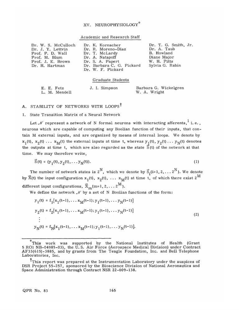
XV. NEUROPHYSIOLOGY* Academic and Research Staff Dr. Dr. Dr. Dr. Dr. Dr. Dr. Dr. W. S. McCulloch Dr. J. Y. Lettvin Prof. P. D. Wall Prof. M. Blum Prof. J. E. Brown Dr. H. Hartman K. Kornacher R. Moreno-Diaz T. McLardy A. Natapoff S. A. Papert Barbara C. G. Pickard W. F. Pickard Dr. T. G. Smith, Jr. Dr. A. Taub B. Howland Diane Major W. H. Pitts Sylvia G. Rabin Graduate Students Barbara G. Wickelgren W. A. Wright J. I. Simpson E. E. Fetz L. M. Mendell A. STABILITY OF NETWORKS WITH LOOPSt 1. State Transition Matrix of a Neural Network Let XA represent a network of N formal neurons with interacting afferents, i. e., neurons which are capable of computing any Boolian function of their inputs, that contain M external inputs, and are organized by means of internal loops. xl(t), x 2 (t) .. . xM(t) the external inputs at time t, whereas Yl(t), Y 2 (t) ... the outputs at time t, time. We denote by YN(t) denotes which are also regarded as the state S (t) of the network at that We may therefore write, S (t) = (y l (t), YZ(t), ... YN(t)). (1) The number of network states is 2 N , which we denote by Si(i= 1, 2, ... by X(t) the input configuration x 1 (t), x 2 (t), ... different input configurations, X We define the network J/ (m= 1, 2,... xM(t) at time t, 2M 2 N ). We denote of which there exist 2 ). by a set of N Boolian functions of the form: Y1(t) = fl[xl(t-l),... xM(t-l); yl(t-1),... YN(t-1)] Y2(t)= f2[xl(t-1),... xM(t-1); yl(t-1),... YN(t-1)] YN(t) = fN[xl(t-1),.. (2) xM(t-1); yl(t-1),... yN(t-1)]. , of Health (Grant National Institutes This work was supported by the 5 RO1 NB-04985-03), the U. S. Air Force (Aerospace Medical Division) under Contract AF33(615)-3885, and by grants from The Teagle Foundation, Inc. and Bell Telephone Laboratories, Inc. TThis report was prepared at the Instrumentation Laboratory under the auspices of DSR Project 55-257, sponsored by the Bioscience Division of National Aeronautics and Space Administration through Contract NSR 22-009-138. QPR No. 83 165 (XV. NEUROPHYSIOLOGY) Equations 2 may be written as Yl(t) = f 1 [X(t-1); S(t-1)] y 2 (t) = f 2 [X(t-1); S(t-1)] (3) YN(t) = fN[X(t-1); S(t-1)]. For a particular configuration, of Y 1 of the inputs, Eqs. 3 are Boolian functions i. e., YN' 2 , ... Xm, y (t) = fl[Xm; S(t-1)] y 2 (t)= f 2 [Xm; S(t-1)] (4) YN(t) = fN[Xm; S(t-l)]. For each value of S(t-1) = Si we obtain a new state (yl(t), 2 (t), ... YN(t)) = S.. For each input X = Xm' Nwe wish to consider the state transition matrix /(X ), i. e. , the mN m Boolian matrix of 2 rows and columns in which the .#(X m)ij term is 1 if the network goes from the state S. to the state S. under the input Xm, and 0 otherwise. these matrices .(Xm) ponents of Sj, have one and only one 1 in each row. Therefore, If (a, P,... v) are the com- i. e. , they constitute the string of zeros and ones that define Sj, -W(X)ij may be written X)ij Si f ) 2( ; Si) ' f i) (5) following the convention in which i fn X fn(; (negation) Si) (6) f (X; S) = f (X; Si). Example 1. Consider the network of Fig. XV-1, given by f1 = xl1 f2 = 2 + x 1Y Y1 2 + x 1Y 1 2 X 2 Y1 + x 2 Y1' The state transition matrix, QPR No. 83 A(X), is 166 for which the functions fl and f2 are YI Y2 Fig. XV-1. Fig. XV-2. 22 x3 Fig. XV-3. QPR No. 83 167 (XV. NEUROPHYSIOLOGY) S. 00 00 01 10 X1X2 x1x2 X1X2 11 01 X1X2 x1x2 X1X2 10 x1x x1X 2 X1x S. 1 11 2 x2 0 0 o) 0 0 0 1 00 1 0 0 2 1x2 X1X 2 0 For example, the input X m = (0, 0), O X1X 2 0 gives _/(0, 0), 1 0 which means that, under the input (0, 0), the transitions of states are (0, 0) -(0, 2. 0) (0, 1) - (1, 0) (1, 0) - (0, 1) (1,1) - (0, 1) Stability and Oscillations Definition 1. A network .V is stable under a constant input, Xim if, under that input, the network, after changing, or not to a new state, will remain in said state regardless of its initial state. Otherwise, the network is said to be unstable under Xm Definition 2. A network is completely unstable under Xm if it is unstable for any given initial state. Definition 3. If a network is unstable under Xm, it oscillates in one or more modes depending upon the state of the network when X was applied. The order of a mode of 2 m oscillation is the number of states which are involved in the oscillation. The conditions of stability for IV/-networks may be derived from their transition state matrices, A'(X m), by using the following algorithm (which is a consequence of the meaning of -(Xm); the proof of it is rather self-evident and has been left as an exercise for the reader): a.) If all terms in the diagonal of f(Xm) are zero, the network is completely unstable, where the converse also holds true. Therefore, the necessary and sufficient condition for complete unstability is that the equation QPR No. 83 168 (XV. ()ii S NEUROPHYSIOLOGY) (7) 0 i has solutions. Under these solutions the network will be completely unstable. Example 2. 1, For the network of Example the inputs which provoke complete instability are the solutions of + X1X2 + 0 = x1 + x2 = 0 + X1X2 x1x2 which has the unique solution x 1 = 1, x2 = 0, i. e. , under the input (1,0) the network is completely unstable. b.) If any terms in the diagonal of '(Xm) are 1, we delete the rows and columns which correspond to them, ending in a new matrix in which two alternatives are possible; bl. Some rows have only O's. b Z . All rows have l's. Under the latter case, the network is unstable when the initial state is any of those states which are present in the reduced matrix. In the former case we delete rows and columns corresponding to the states whose rows are all zeros. A new matrix is obtained that follows either alternatives b 1 or b Z . If it follows bl, we continue the process of reduction until we end in a minor that follows b 2 . If, by iteratively applying bl, we end in only one state, the network is stable. Example 3. (0, 0)= /(0, The matrix 0 o 1 0o 0 0 1 0 0 (0 0) for the network of Fig. XV-1 is 1 0 By deleting the first row and column, which have 1 in the diagonal, we obtain ' (0,0)= 0 0 1 which follows b2 . Therefore, under the input (0, 0), the network is unstable if the initial state is either (0,1), (1,0), or (1, 1). W(1, 1) is For the same network, 0 0 1 (1 1)0 0 1 0 0 S 1 0 0 0 0 0 0 1 By deleting the second row and column, we conclude that the network is unstable under the input (1,1) if the initial state is either (0,0), (1,0), or (1,1). QPR No. 83 169 Similarly, the network (XV. NEUROPHYSIOLOGY) is unstable for the input (0, 1) if the initial state is either (0, 0), Example 4. neuron is, 1 YY 3 f2 Yl 2 = The network of Fig. XV-2 has no external inputs. or (1, 1). The function of each respectively, fl f3 (0, 1), Y2Y3 + Y1Y3 + 2Y3 The state transition matrix is (blanks are zeros) (fl'f 1 (Yl,Y 2 , Y3) 1 000 2 001 3 010 4 011 5 100 6 101 7 110 8 111 2 3 4 2 f 3) 5 6 7 8 1 1 1 1 1 1 1 1 By inspection of the diagonal, we can delete rows and columns 1 and 5. Applying criterion bl, we then delete 8 and 4. Reapplying criterion bl, we delete 2; again, we delete 3 and 6, ending with a single state, the 7 th Therefore, the network is stable for any given initial state. 3. Stability for a Single Neuron For the case of a single neuron computing any of the possible Boolian functions of its M inputs, the necessary and sufficient conditions for stability adopt a much simpler form. In this case, we have only one Boolian function that describes the neuron, which is of the form: y(t) = f[X(t-l);y(t-l)] (8) The transition state matrix, .4(X), is (X)= QPR No. 83 f(X; 0) f(X, 0) f(X; 1) f(X; 1) 170 (XV. NEUROPHYSIOLOGY) Since there exist only two states, unstability of any kind implies complete unstability. Therefore, the necessary and sufficient conditions for stability are manifest in stipulating that the equation (10) f(X; 0) + f(X; 1) = 0 has no solution, i. e., the solutions of Eq. 10 produce unstability. neuron will be characterized by the simplest oscillation 010101... By negating Eq. 10, we obtain If unstable, the (11) f(X; 0) • f(X; 1) = 1 Therefore, the solutions of (12) f(X; 0) • f(X; 1) = 0 are inputs under which the neuron is stable, i. e. , Eq. 12 gives the necessary and sufficient condition for stability. Example 5. Consider the neuron of Fig. XV-3, which computes the function f = x 1 x2 x 3Y + (x 1+xz)y. Equation 12 now takes the form X1X2x3 • (X 1 +x 2 ) = x 1 x 2 x 3 x 1 x 2 = 0. Therefore, the neuron is stable for any input. R. Moreno-Diaz References 1. 2. B. W. S. McCulloch, "Agathe Tyche of Nervous Nets - The Lucky Reckoners," in Embodiments of Mind (The M. I. T. Press, Cambridge, Massachusetts, 1965). R. Moreno-Diaz, "Realizability of a Neural Net Capable of All Possible Modes of Oscillation," Quarterly Progress Report No. 82, Research Laboratory of Electronics, M.I.T., July 15, 1966, pp. 280-285. MYELINATED FIBERS IN THE DORSAL ROOTS OF CATS With Dr. J. Y. Lettvin we have been studying the electrical properties of those structures in cat intradural dorsal root which we could impale with KC1-filled micropipettes. The experimental technique and preliminary results were reported in Quarterly Progress Report No. 78 (pages 281-282). We report here the conclusions reached after using two different impalement techniques. QPR No. 83 171 (XV. NEUROPHYSIOLOGY) In the first technique (tapped penetrations), the micropipette tip was positioned in a region of high resistivity by using a micrometer and the end of the micrometer barrel tapped. This normally resulted either in returning the tip to a region of low resistivity and no resting potential or in penetrating a unit showing approximately -70 my resting potential, electrotonic charging properties, and, when sufficiently stimulated, a fullscale (>80 my) action potential. Since the shape of the electrotonic charging curve was consistent with that expected for a parallel RC circuit, and since this would not be the case if we were charging some unit through a length of internode, we believe that the RC combination is at the point of penetration into the fiber. Similarly, we believe the point of penetration to be a point of excitability because, if it were not, the RC combination would give rise to a shunting of the action potentials from neighboring excitable regions, and this was not noticed. able membrane. Hence, at the point of penetration there is an excit- That this membrane is not associated with a naturally occurring node of Ranvier can be inferred from the fact that the observed resistances (R~10 M) much too low and the observed capacitances (C~6 pf) much too high, are which the fre- quency of penetration is much higher than would be expected if only Ranvier nodes could be penetrated. We conclude, therefore, that penetration must be internodal and that the act of penetration creates an artificial node of Ranvier whose somewhat larger than that of naturally occurring ones. area is We account for this by sup- posing that tapping of the micrometer barrel causes a complex orbital motion of the micropipette tip which eventually ruptures the myelin, leaving the way open for penetration of the axolemma which must be excitable along its entire length. W. F. C. Pickard FURTHER STUDIES WITH CYLINDER LENSES In Quarterly Progress Report No. 73 (pages 309-316), we have described a method for testing camera lenses which makes use of the properties of the crossed-cylinder lens. In a later report (Quarterly Progress Report No. 77, pages 383-389), we showed how a small section of such a lens can be fabricated by the torsional deformation of a prism of glass. It now appears that this method can be generalized to permit the fabri- cation of a large-aperture sphero-cylinder lens. The method depends on the deformation of square plates by couples of force at opposite corners. The treatment of this problem in elasticity theory may be found in standard texts; a formula that is valid for deformations that are small compared with the thickness of the plates is given by l , 2 z = k x y. (1) We change coordinates as follows: QPR No. 83 172 (XV. 1 a = NEUROPHYSIOLOGY) (x+y) 1 p 1 (x-y), and note that x 2 2 +y 2 2 =a +p= r 2 Equation 1 may be transformed as follows: z =k x z =- k2r y [x2+y -(x-y)2] -kP 2 The first and second terms of Eq. 2 represent, respectively, a spherical component of curvature k/2, and a cylinder component with axes a, p and curvature -k. This par- ticular combination of spherical and cylinder components is known as the "crossedcylinder lens," and is used extensively in ophthalmic refractive diagnosis.3 If one wishes to isolate the "pure" cylinder component, corresponding to the second term of Eq. 2, an additional spherical lens may be employed to cancel the spherical component. The feature of this cylinder lens construction that recommends it in preference to more obvious methods, as for example, wrapping a sheet of glass around a precisely formed cylindrical mandrel of large radius of curvature, is that the forces are selfequalized and need be applied at only four points. 1. Practical Features in the Construction of Such a Lens It will be apparent that there is a maximum refractive power, relative to the size of the plates, which can in practice be attained because of the combination of two limitations: the deformation of the plate must be small compared with the thickness of the plate, in order that Eq. 1 will apply, and the breaking strength of the glass must not be exceeded. (Quartz would give greater strength; various plastics would permit greater deformation before breaking but would have inferior optical properties.) Let us now consider the various methods by which cylinder lenses can be fabricated from deformed plates of glass. In Fig. XV-4 we show two plates, each 3 mm thick and 78 mm square, which have been spaced with rubber shims, those in one pair of opposite corners being twice the thickness of the others, and taped to provide a reservoir for fluid or plastic. (With careful manipulation, the tape can be applied after the glass has been deformed by tightening the clamps.) The space between the plates may be filled with an inert transparent fluid of low vapor pressure such as a silicone oil, or it can be QPR No. 83 173 (XV. NEUROPHYSIOLOGY) cast with an epoxy resin for a more permanent and useful construction. For this prupose, we can recommend Maraset Type 657 A and B casting resin combination (from The Fig. XV-4. Two square optical glass plates, taped to contain liquid or epoxy resin, are stressed in torsion by external clamps and internal rubber shims. Marblette Corporation, Long Island City, N. Y.) mixed as directed, centrifuged to remove bubbles, and allowed to harden undisturbed at room temperature for one week. Two out of six lenses fabricated in this way have been judged to be near-perfect; method is still subject to some variability. the Useful results have been obtained with these plastic-cast lenses, but there are two evident disadvantages to this construction: the glass is left under strain and is thus prone to fatigue failure or breakage with small shocks; and the homogeneity of the epoxy plastic is inferior to that of optical glass, and is liable to change with absorption of solvents, age, heat, and so forth. A way around these difficulties is suggested by a study of the method used by Bernhard Schmidt to fabricate the corrector plate for his famous tele4 scope design. This method of construction, which we have not yet tested, is the following: One starts with the previous lens, epoxy resin cast between the deformed glass plates. This sandwich of glass and plastic is ground and polished flat on the outer surfaces, and the components are then separated by using heat, solvents or both. These glass components are each crossed-cylinder lenses of dioptric strength determined by the index of the glass rather than the plastic. They may be used separately, or may be glued together at their now flat inner surfaces by using the thinnest possible layer of cement, to form a double-strength, unstrained, cross-cylinder lens of superior quality. QPR No. 83 174 (XV. 2. NEUROPHYSIOLOGY) Tests of Plastic-Cast Crossed-Cylinder Lens The method that we use for testing crossed-cylinder lenses is the following: A collimator is first adjusted to project an image of a monochromatic point source. This simulated monochromatic star image is photographed with a long focal length camera lens of aperture similar to that of the collimator. Interposed between these two lens Fig. XV-5. Test of crossed-cylinder lens of new construction; space between plates has been cast in epoxy resin. A perfect lens would image the grid without distortion. systems is the crossed-cylinder lens, together with a rectangular grid, here a photochemically etched screen of beryllium copper having a 'wire' thickness that is small compared with the mesh spacing, which is 2-5 mm. The grid is oriented so as to give minimal diffraction blur, which means that its axes are parallel to the edges of the deformed square plate lens. With this arrangement, the camera photographs what is in effect an image in miniature of the aperture of the cylinder lens as metered by the grid. The quality of the cylinder lens is indicated by the degree of distortion of the image of the rectangular grid. In Fig. XV-5 we show the results of a test of a plastic-cast lens of 78-mm square aper[The optician's formula for this lens is (-1/8, ture, and strength ±1/8 diopter. +1/4 Diopters).] For this test, the angular subtense of the collimator was 1/50,000 radian, and the spectrum of the source was restricted to 5750 ± 50 A; the camera and collimator lenses were high-quality air-spaced doublet lenses. Thus, we can reasonably ascribe the defects in the pattern of this grid, which is seen to exhibit a 'folding over' at the corners where the external clamps were applied, to the cylinder-lens construction. The QPR No. 83 175 Fig. XV-6. Comparison of crossed-cylinder lenses of same dioptric powers and differing constructions. (a) Lens of new construction. (b) Ophthalmic lens. 4 'Ass X # sixa Fig. XV-7. Distortion of the pattern of the grid caused by spherical aberration. QPR No. 83 Fig. XV-8. 176 Distortion of the pattern of the grid caused by coma. (XV. NEUROPHYSIOLOGY) central regions of this lens (not our best lens) are, however, unaffected by this defect. Lenses of this type were fabricated from very good quality plate glass of 2. 5-3 mm thickness; this power is the highest possible without probable breakage caused by delayed fatigue failure of the glass. By coincidence, this is also the smallest available prescription power available in ophthalmic crossed-cylinder lenses, of optical quality. and permits a comparison In Fig. XV-6a we show a test on a finer grid than before, central 50 mm of our very best plastic-cast cylinder lens. In Fig. XV-6b is similar test of the best of three ophthalmic crossed-cylinder lenses. formance of the new construction is thus demonstrated, obviously an unfair one. of the shown a The improved per- although the comparison is The defects of the ophthalmic lens shown here are so small that they could not be observed by any test with an unaided human eye. 3. Use of Crossed-Cylinder Lens to Test Camera Lenses The method that we use to test crossed-cylinder lenses can also serve as a test of the camera lens, once the quality of the crossed-cylinder lens has been established. The distortion of the pattern of the grid will give qualitative indication of the predominate aberration of the lens. In Fig. XV-7 we show the pattern of the grid characteristic of primary spherical aberration, simulated here by means of a supplementary lens pair, a flat convex and a bent concave lens of opposite dioptric powers. In Fig. XV-8 we show the distortion characteristic of the off-axis aberration coma. We regard this extension of our previously reported lens-testing procedures as being of importance, aperture. since it enables one to test the performance of the lens over its entire Thus one can detect defects of figuring, as well as formula. possibility is to test optical elements, for example, Still another mirrors, prisms, filters, window glass elements, thereby using the instrument in the manner of a striascope. The advantage is that one need not record or interpret light-intensity variations; the distortion of the pattern of the grid gives the information. An advantage of methods that use the crossed-cylinder lens over other lens tests that make use of aperture screens (of which the Hartmann test is perhaps the best example) is that the axis of the grid can be adjusted so as to give minimal diffraction blurring of the pattern. Thus, only to the extent that the lens produces aberrations affecting the orientation of the elements of the rectangular grid, is the pattern subject to diffraction blurring. In effect, then, the more perfect the lens, or other element under test, the more sensitive is the test. In our present experiments the brightness and spot size of our collimator limit the magnification that can be attained with time exposures of reasonable duration. A neon laser of modest power should remove this limitation and enable us to determine the ultimate sensitivity of the method. B. Howland, S. J. [Stephen J. Wiesner is with the Department of Physics, Brandeis University.] QPR No. 83 177 Wiesner (XV. NEUROPHYSIOLOGY) References 1. A. E. H. Love, A Treatise on Mathematical Elasticity New York, n. d.), p. 471. 2. S. Timoshenko and S. Woinowsky-Krieger, Theory of Plates and Shells Hill Book Company, New York, 1959), Chap. 2. 3. I. M. Borish, Clinical Refraction (The Professional Press Inc. , Chiago, Illinois, 1954). 4. Amateur Telescope Making, Book Three, Albert G. Ingalls (ed.)(Scientific American, Publisher, New York, 1961), p. 365. QPR No. 83 178 (Dover Publications, (McGraw-


