INSIDE -The new Interprofessional Dizziness and Balance Clinic: p. 5 pacificu.edu
advertisement
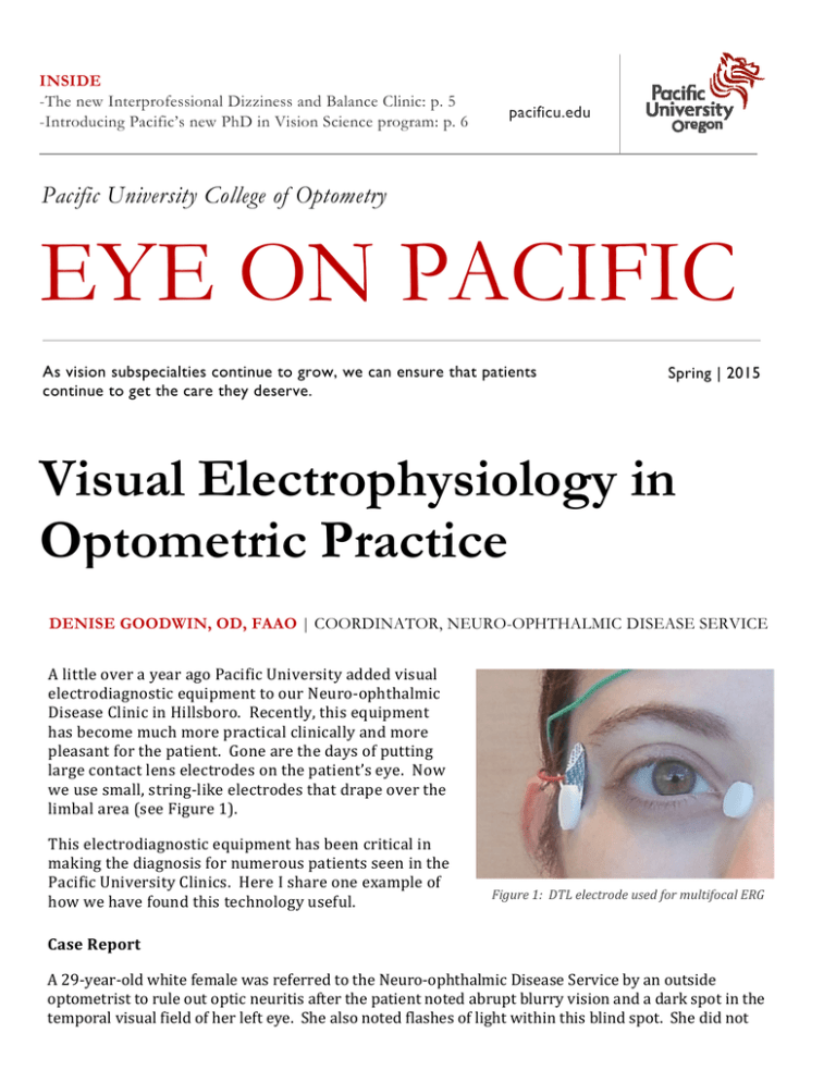
INSIDE -The new Interprofessional Dizziness and Balance Clinic: p. 5 -Introducing Pacific’s new PhD in Vision Science program: p. 6 pacificu.edu Pacific University College of Optometry EYE ON PACIFIC As vision subspecialties continue to grow, we can ensure that patients continue to get the care they deserve. Spring | 2015 Visual Electrophysiology in Optometric Practice DENISE GOODWIN, OD, FAAO | COORDINATOR, NEURO-OPHTHALMIC DISEASE SERVICE A little over a year ago Pacific University added visual electrodiagnostic equipment to our Neuro-ophthalmic Disease Clinic in Hillsboro. Recently, this equipment has become much more practical clinically and more pleasant for the patient. Gone are the days of putting large contact lens electrodes on the patient’s eye. Now we use small, string-like electrodes that drape over the limbal area (see Figure 1). This electrodiagnostic equipment has been critical in making the diagnosis for numerous patients seen in the Pacific University Clinics. Here I share one example of how we have found this technology useful. Figure 1: DTL electrode used for multifocal ERG Case Report A 29-year-old white female was referred to the Neuro-ophthalmic Disease Service by an outside optometrist to rule out optic neuritis after the patient noted abrupt blurry vision and a dark spot in the temporal visual field of her left eye. She also noted flashes of light within this blind spot. She did not Visual Electrophysiology (continued) report pain with eye movements, diplopia, dizziness, fever, or nausea or vomiting, but she did say that both feet became numb at night. She was not taking any medications. Family history was noncontributory. Best corrected visual acuities were 20/20 in the Figure 2: Fundus photos demonstrating mild edema in the left eye right and 20/30 in the left eye. Pupils were round and reactive to light, with no relative afferent optic nerve appeared round with distinct margins. pupillary defect (RAPD). Color vision with HHR There were no hemorrhages or cotton wool spots pseudoisochromatic plates was 10/10 in the right in either eye. The macula was flat and dry in each eye and 6/10 in the left eye, with 80% red eye. desaturation in the left eye. There was slightly Consistent with the fundus photos, optical reduced hearing on the right ear. Cranial nerves coherence tomography demonstrated mild 5, 7, 9, 10, 11, and 12 were intact bilaterally. thickening of the nerve fiber layer inferiorly in the The anterior segment of both eyes was left eye (Figure 3). A 30-2 Humphrey Visual Field showed an absolute scotoma surrounding the unremarkable. Intraocular pressures with Goldmann applanation tonometry were 15 mmHg physiologic blind spot of the left eye (Figure 4). in each eye. Examination of the posterior Given her age, race, gender, and mild unilateral segment revealed an anomalous optic nerve with disc edema, we, like the referring optometrist, slightly blurry disc margins, particularly suspected the patient had optic neuritis. inferiorly, in the left eye (Figure 2). The right Therefore, we ordered an MRI of the orbits and brain with contrast. However, the neuroimaging did not show any evidence of demyelinating plaques or inflammation of the optic nerve. Figure 3: Nerve fiber layer OCT showing mild edema of the nerve fiber layer inferiorly Since the MRI ruled out most optic neuropathies, we decided to rule out an occult retinal cause by performing a multifocal electroretinogram (ERG). Here, the left eye showed significant depression of the nasal retinal response that corresponded to the visual field defect (Figure 5). This helped us isolate the exact anatomical location of the disease process and helped us make the diagnosis. The abnormal ERG indicated that the problem was in the retina, specifically in the outer retinal layers. This, combined with her demographics, signs, and symptoms, led us to diagnose acute idiopathic blind spot enlargement (AIBSE). Visual Electrophysiology (continued) Discussion AIBSE is a condition of unknown origin that affects the outer retinal layers of the peripapillary retina. It occurs most commonly in women aged 16 to 50 years. Photopsia, followed by vision loss, are the most common visual symptoms. Visual acuity may be affected, but severe acuity loss is unusual. Dyschromatopsia is a common finding, and a relative afferent pupillary defect may be present. All patients have an enlarged blind spot. The scotoma is variable in size but is usually absolute with steep borders. Patients with AIBSE may have mild optic nerve swelling, as well as peripapillary pigment changes. Occasionally there are multiple white retinal lesions, similar to that seen with multiple evanescent white dot syndrome (MEWDS). Photopsias and visual acuity resolve without treatment, but visual field and ERG defects often persist indefinitely. Recurrences can occur. In this case, the patient regained 20/20 acuity in the left eye, and the photopsias ceased. The visual field defect remains stable. AIBSE is frequently misdiagnosed as optic neuritis due to the demographics and presentation. It can also be mistaken for an ocular migraine due to the presence of photopsias in a relatively normal appearing fundus. Papilledema can result in an enlarged blind spot, but the edema associated with AIBSE is mild relative to the size of the scotoma. AISBE must also be differentiated from a chiasmal lesion that can cause temporal visual field defects. In this case, the multifocal ERG was critical in making the diagnosis. Figure 4: Visual fields showing an enlarged blind spot in the left visual field. Figure 5: Multifocal ERG showing decreased retinal function surrounding the optic nerve of the left eye. vision loss. Other conditions in which the ERG may help with the diagnosis include occult macular dystrophy, hydroxychloroquine toxicity, and cancer associated retinopathy. The multifocal ERG measures the response of the outer retina, the cones and bipolar cells. The multifocal ERG defect should correlate with the visual field defect; otherwise, an alternative diagnosis should be considered. A combination of ERG and visual evoked potential (VEP) testing can be particularly valuable in distinguishing between retinal and neurologic conditions, as well as determining the cause of unexplained vision loss. The VEP is also valuable in managing glaucoma and other optic neuropathies. The most common reason we perform electrodiagnostic testing in our Hillsboro Clinic is to help determine the cause of unexplained Please contact the Hillsboro Clinic at 503-352-7300 if we can serve you and your patients in our Neuroophthalmic Disease and Electrodiagnostics Service. Advances in Contact Lenses FRANK ZHENG, OD | CONTACT LENS RESIDENT In many western cultures, making eye contact is a fundamental social skill. Individuals with iris defects or visible blemishes may experience feelings of being self-conscious or embarrassment. Fortunately for such patients, ocular injury or iris defects can be successfully disguised with prosthetic contact lenses. Made in a variety of refractive powers, transmissible tints, and iris designs, these lenses can be made to perfectly match the patient’s visual and cosmetic needs. Beyond cosmetic fixes, such lenses may also be used as a management option for patients with photophobia, iris anomalies, albinism, strabismus, or amblyopia. The members of the therapeutic contact lens team at Pacific University include Pat Caroline; Mark Andre; Beth Kinoshita, OD; Matthew Lampa, OD; and our current Cornea and Contact Lens Resident, Frank Zheng, OD. If you have patients who may require such specialty contact lens care we would be honored to serve them in our Therapeutic Contact Lens Clinic. Advances in Medical Eye Care LORNE YUDCOVITCH, OD, MS, FAAO | MEDICAL EYE CARE SERVICE CHIEF A recommendation I like to give interns when they start seeing patients is: Never touch a ‘red eye’. Specifically, if there is ocular surface compromise, and physical contact does not provide better diagnosis or treatment (and may compromise ocular health further), non-invasive tests are preferred. ‘Red eye’ in this case refers to narrow angles; dislocated intraocular lenses; wound leaks; corneal abrasions, lacerations, or thinning; or other conditions that compromise corneal integrity. The patient may have a contraindication to anesthetic use, decline invasive testing, have special needs, or be in an age range that makes contact testing difficult. Anterior segment optical coherence tomography (OCT) often allows us to bypass the need for anesthetics or testing that involves contact with the ocular surface. Anterior chamber angle assessment can be performed in any of the 360 Anterior segment evaluation with the Visante OCT Optovue pachymetry map degrees, helping to confirm or rule out narrow angles. Corneal disease, iris cysts, synechiae, iridotomy, bleb patency, and intraocular lens position are other examples that can be evaluated non-invasively. Pachymetry is possible without using the traditional contact probe, often providing more precise measurements. We are happy to consult with you and/or accept referrals for anterior segment imaging. Advances in Binocular Vision BRADLEY COFFEY, OD, FAAO | INTERPROFESSIONAL DIZZINESS AND BALANCE CLINIC The NEW Pacific University Interprofessional Dizziness and Balance Clinic is a diagnostic clinic dedicated to improving the lives of our patients by sending them on the proper path to recovery. This clinic’s unique approach capitalizes on thorough testing of the three major systems for balance; vestibular, visual, and proprioceptive; to determine the cause of the imbalance and recommend the best treatment modalities. The concept for this clinic has been simmering for a few years and was brought to fruition by the arrival of Annie Hogan, PhD, a faculty member in the School of Audiology with expertise in vestibular function. Dr. Hogan recruited Katie Farrell, PT, DSc, and Bradley Coffey, OD as the founding faculty for the Balance Clinic. They see Balance Clinic patients directly while interns from each discipline observe the team in action. Dr. Hogan’s testing focuses on the vestibular system, utilizing videonystagmography, as well as cervical and ocular vestibular-evoked myogenic potentials to get a better understanding of the role of the vestibular system plays in the patient’s symptomatology. Dr. Farrell’s testing focuses on the somatosensory and physical performance aspects of our balance system to determine if there are impairments that contribute to an individual’s imbalance. Dr. Coffey’s testing evaluates the oculomotor, dynamic visual, and visual-vestibular systems to determine the extent these systems play into the patient’s symptoms. Patients are seen in the Balance Clinic after obtaining primary care vision and hearing tests either from community providers or from the University Clinics. On the day of the appointment, each patient is examined by each Balance Clinic clinician, after which the faculty and interns gather to discuss each case and recommend treatment or further assessment. It is not the individual clinicians that make this clinic unique, but rather the combined, collaborative expertise and analysis that allows us to best understand the patient as a whole. With a thorough understanding of the balance system, and its areas of dysfunction, we can guide the patient to the best treatment modalities for a faster recovery. If you would like to arrange a referral to the Interprofessional Dizziness and Balance Clinic, please contact the Pacific EarClinic by phone 503352-2692 or by email at earclinic@pacificu.edu. Advances in Pediatric Eye Care JP LOWERY, OD, FAAO | PEDIATRIC VISION SERVICE CHIEF Most of us have auto-refractors (AR) in our clinics and we all use them as a starting point for refraction in adults. Since we cannot get reliable subjective refractions on preschoolers, the AR may be an alluring option for objective findings. It is usually possible to obtain AR readings with young children over age 2, but should we trust these measures? A wealth of research and experience has shown that standard ARs do not control accommodation in young children. The "fogging" mechanisms encourage the child to accommodate, resulting in “instrument myopia.” Therefore, the spherical component of the refractive measurement is only valid when the child is cyclopleged. The cylinder power is valid with or without cycloplegia. In fact, due to variations in accommodative posture over time, the "dry" auto-refraction value is often more accurate than the dry retinoscopy cylinder value simply because the AR is able to measure both primary meridians simultaneously while we are measuring each meridian separately when we perform sphere-sphere retinoscopy. I find the AR is especially helpful for determining an accurate cylinder axis with young children, particularly in those with high amounts of amblyogenic astigmatism. It can be difficult to determine the axis with streak retinoscopy so if I get multiple AR readings that are all within 5 degrees, I feel much more confident writing my final prescription. Stay tuned for the upcoming series on prescribing tips in pediatric eye care. Advances in Eye Care Research Yu-Chi Tai, PhD | VISION SCIENCE GRADUATE PROGRAM DIRECTOR Accredited on March 6, 2015, Pacific University announces its new PhD in vision science (PhDVS) program and becomes the first private institution offering a PhD degree in the field. Joining Pacific’s highly regarded doctor of optometry (OD) and the blooming master in vision science (MSVS) programs, the new PhD program provides students with comprehensive knowledge in contemporary vision science and quality training though rigorous hands-on laboratory/research/teaching experiences, equipping them to be professional vision scientists and/or educators. The program also supports cutting-edge research in basic, clinical, and translational vision research. Students study with faculty whose interests span a broad range of specialty areas. They also collaborate and network with scientists and/or industry around the world through research projects. Students with prior education from many related disciplines are accepted. Applications are accepted September 1 through March 15. Detailed information can be found at: http://pacificu.edu/optometry. Faculty in the News CAROLE TIMPONE, OD, FAAO, FNAP | ASSOCIATE DEAN OF CLINICAL PROGRAMS Through the College of Optometry’s Outreach Program, Pacific University reaches out to our community providing mobile eye exams on site at vineyards, convention centers, schools, churches, health fairs, and other locations where the underserved are in need of services. You may have seen Dr. Sarah Martin, driving the Pacific University College of Optometry EyeVan on her way to one of these community service events. I am pleased to announce that Dr. Martin, recently awarded a Pacific University Faculty Development Grant for “Enhancement and Expansion of the Community Outreach Program,” has been named the College’s new Director of Community Outreach. Coming to Pacific in 2013, Dr. Martin is originally from Southern California and a 2007 graduate of New England College of Optometry. She completed a residency in Primary Care and Low Vision at the Southern Arizona VA Hospital in Tucson, followed by three years in the Indian Health Service where she helped establish a remote eye clinic. Now Dr. Martin enthusiastically supervises students as they perform vision screenings and mobile eye exams. Sarah is very passionate about public health and education and is honored to lead the community outreach program. In her spare time she enjoys tennis, hiking, swimming, and traveling with her family. Sue Littlefield, OD, a Pacific University College of Optometry alumna, served as the college’s Director of Community Outreach for 10 years. In July, Dr. Littlefield is looking forward to assuming her new role as Director of Pacific’s Beaverton EyeClinic. As Director of Community Outreach, she successfully developed and enhanced the outreach program, making it more prominent in the curriculum. She had several years’ experience running her private practice in Seattle, before returning to Pacific to join the faculty. Most recently, she has been Clinic Director of Pacific’s EyeClinic in the Virginia Garcia Health and Wellness Center in Cornelius. Susan is committed to enhancing community collaborations and partnerships, and developing student, staff and attending doctors’ ability to implement evidence based leading edge vision care. Dr. Littlefield’s scholarly interests and grant awards have included studies in health disparities, community based education, and projects involving underserved populations. In addition to her duties as Clinic Director, she teaches Patient Care and Refractive Surgery courses. We also recognize and thank Dr. Ami Halvorson, founding Director of the Beaverton EyeClinic, for her years of service, as she begins her new position at Pacific Cataract and Laser Institute. Our clinical faculty are here to help you with patient consultations and referrals. Please let us know how we can best serve your needs! CE Opportunities Research Opportunities April: -PUCO Coeur d’Alene CE; Coeur d’Alene Golf and Spa Resort; April 24-25 -College of Health Professions – Deciphering the Complex Nature of Balance and Vestibular Dysfunction; Pacific Health Professions Campus, Hillsboro, OR; April 25 We are recruiting patients for two National Eye Institute sponsored studies. The Convergence Insufficiency Treatment Study is evaluating the effectiveness of home-based therapy for symptomatic convergence insufficiency. Children should be 9 to <18 years of age with symptomatic convergence insufficiency. The Hyperopic Treatment Study is evaluating when to prescribe for hyperopia in very young children. We are recruiting hyperopes +3.00 to +6.00. There are 2 Cohorts: children 12 months to <36 months of age and 36 to <72 months of age. For recruitment criteria or to refer a candidate, please contact study coordinator, Jayne Silver, at jaynesilver@comcast.net or 503-804-8241. May: -Teplick 20th Annual Blockbuster CE; Hotel deluxe, Portland, OR; May 30 June: -PUCO Residents Conference; Jefferson Hall, Forest Grove, OR; June 12-13 July: -Victoria Conference; Inn at Laurel Point, British Columbia, Canada; July 16-19 -OOPA Summer CE Event; The Resort at the Mountain, Welches, OR; July 16-18 Practice Management Tips BARBARA COLTER | DIRECTOR OF CLINICAL OPERATIONS Start wrapping your mind around glaucoma coding. As you know, the goal of ICD-10 is to pack as much information as possible into a single diagnosis code. For instance, effective Oct. 1, 2015, about 250 codes will be available from which to select when coding for glaucoma. Here is an example: We will code a severe case of glaucoma associated with ocular trauma to the left eye as H40.32X3. The corresponding ICD9 code would be 365.65. Characters 1-3 indicate the general condition. The codes H00-H059 represent diseases of the eye and adnexa. In this case, H40 is for glaucoma. Character 4 indicates the type of the condition. In our example, H40.3 indicates that the glaucoma is secondary to eye trauma. Character 5 indicates the location: 1 - right eye, 2 left eye, 3 - bilateral, 9 -unspecified eye. Character 6 is an X. This is specified as a place holder for future needs. Character 7 describes the severity of the condition: 1 - mild, 2 - moderate, 3. - severe, 4 - indeterminate. Not all diagnosis codes will have seven characters. It is important to learn those that will have a place holder and those that will not. Bottom line: start memorizing the numbers for location and severity. They are the same across the board. It is also a great idea to find a source that will readily assist you when looking up these codes. There are many great online options if your EHR is not sufficient. Referral Service Contact Numbers Pacific EyeClinic Forest Grove 2043 College Way, Forest Grove, OR 97116 Phone: 503-352-2020 Fax: 503-352-2261 Vision Therapy: Scott Cooper, OD; Graham Erickson, OD; Hannu Laukkanen, OD; JP Lowery, OD Pediatrics: Scott Cooper, OD; Graham Erickson, OD; Hannu Laukkanen, OD; JP Lowery, OD Medical Eye Care: Ryan Bulson, OD; Tracy Doll, OD; Lorne Yudcovitch, OD Low Vision: Karl Citek, OD; JP Lowery, OD Contact Lens: Mark Andre; Tad Buckingham, OD; Patrick Caroline; Beth Kinoshita, OD; Scott Pike, OD Pacific EyeClinic Cornelius 1151 N. Adair, Suite 104 Cornelius, OR 97113 Phone: 503-352-8543 Fax: 503-352-8535 Pediatrics: JP Lowery, OD Medical Eye Care: Tad Buckingham, OD; Len Koh, OD; Sarah Martin, OD; Lorne Yudcovitch, OD Pacific EyeClinic Hillsboro 222 SE 8th Avenue, Hillsboro, OR 97123 Phone: 503-352-7300 Fax: 503-352-7220 Pediatrics: Ryan Bulson, OD Medical Eye Care: Tracy Doll, OD; Dina Erickson, OD; Len Koh, OD; Caroline Ooley, OD Neuro-ophthalmic Disease: Denise Goodwin, OD Pacific EyeClinic Beaverton 12600 SW Crescent St, Suite 130, Beaverton, OR 97005 Phone: 503-352-1699 Fax: 503-352-1690 3D Vision: James Kundart, OD Pediatrics: Alan Love, OD Medical Eye Care: Drew Aldrich, OD; Susan Littlefield, OD Contact Lens: Matt Lampa, OD Pacific EyeClinic Portland 511 SW 10th Ave., Suite 500, Portland, OR 97205 Phone: 503-352-2500 Fax: 503-352-2523 Vision Therapy: Bradley Coffey, OD; Ben Conway, OD; Scott Cooper, OD; James Kundart, OD Pediatrics: Bradley Coffey, OD; Ben Conway, OD; Scott Cooper, OD; James Kundart, OD Medical Eye Care: Ryan Bulson, OD; Candace Hamel, OD; Scott Overton, OD; Carole Timpone, OD Contact Lens: Mark Andre; Candace Hamel, OD; Matt Lampa, OD; Scott Overton, OD; Sarah Pajot, OD Neuro-ophthalmic Disease/Strabismus: Rick London, OD Low Vision: Scott Overton, OD When scheduling an appointment for your patient, please have the patient’s name, address, phone number, date of birth, and name of insurance, as well as the type of service you would like Pacific University eye clinics to provide.
