MODULATED DUAL SATURATION PULSE TRAINS FOR FAIR STUDIES OF CEREBELLUM...
advertisement

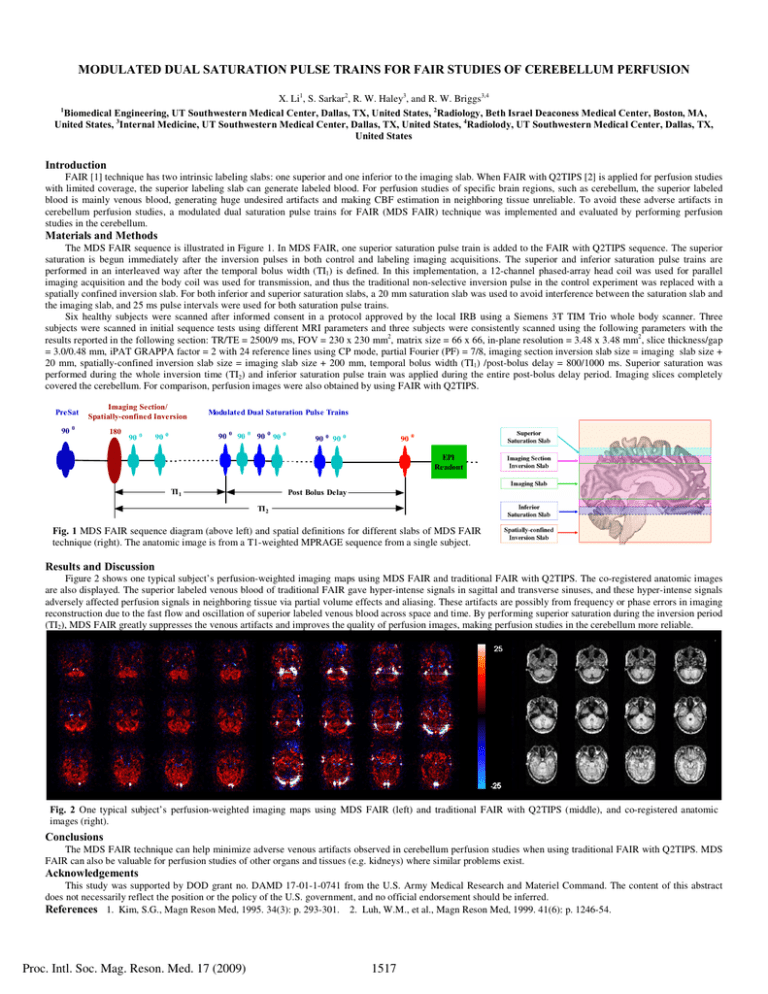
MODULATED DUAL SATURATION PULSE TRAINS FOR FAIR STUDIES OF CEREBELLUM PERFUSION X. Li1, S. Sarkar2, R. W. Haley3, and R. W. Briggs3,4 Biomedical Engineering, UT Southwestern Medical Center, Dallas, TX, United States, 2Radiology, Beth Israel Deaconess Medical Center, Boston, MA, United States, 3Internal Medicine, UT Southwestern Medical Center, Dallas, TX, United States, 4Radiolody, UT Southwestern Medical Center, Dallas, TX, United States 1 Introduction FAIR [1] technique has two intrinsic labeling slabs: one superior and one inferior to the imaging slab. When FAIR with Q2TIPS [2] is applied for perfusion studies with limited coverage, the superior labeling slab can generate labeled blood. For perfusion studies of specific brain regions, such as cerebellum, the superior labeled blood is mainly venous blood, generating huge undesired artifacts and making CBF estimation in neighboring tissue unreliable. To avoid these adverse artifacts in cerebellum perfusion studies, a modulated dual saturation pulse trains for FAIR (MDS FAIR) technique was implemented and evaluated by performing perfusion studies in the cerebellum. Materials and Methods The MDS FAIR sequence is illustrated in Figure 1. In MDS FAIR, one superior saturation pulse train is added to the FAIR with Q2TIPS sequence. The superior saturation is begun immediately after the inversion pulses in both control and labeling imaging acquisitions. The superior and inferior saturation pulse trains are performed in an interleaved way after the temporal bolus width (TI1) is defined. In this implementation, a 12-channel phased-array head coil was used for parallel imaging acquisition and the body coil was used for transmission, and thus the traditional non-selective inversion pulse in the control experiment was replaced with a spatially confined inversion slab. For both inferior and superior saturation slabs, a 20 mm saturation slab was used to avoid interference between the saturation slab and the imaging slab, and 25 ms pulse intervals were used for both saturation pulse trains. Six healthy subjects were scanned after informed consent in a protocol approved by the local IRB using a Siemens 3T TIM Trio whole body scanner. Three subjects were scanned in initial sequence tests using different MRI parameters and three subjects were consistently scanned using the following parameters with the results reported in the following section: TR/TE = 2500/9 ms, FOV = 230 x 230 mm2, matrix size = 66 x 66, in-plane resolution = 3.48 x 3.48 mm2, slice thickness/gap = 3.0/0.48 mm, iPAT GRAPPA factor = 2 with 24 reference lines using CP mode, partial Fourier (PF) = 7/8, imaging section inversion slab size = imaging slab size + 20 mm, spatially-confined inversion slab size = imaging slab size + 200 mm, temporal bolus width (TI1) /post-bolus delay = 800/1000 ms. Superior saturation was performed during the whole inversion time (TI2) and inferior saturation pulse train was applied during the entire post-bolus delay period. Imaging slices completely covered the cerebellum. For comparison, perfusion images were also obtained by using FAIR with Q2TIPS. PreSat 90 Imaging Section/ Spatially-confined Inversion 0 180 90 0 Modulated Dual Saturation Pulse Trains 90 0 90 0 90 0 90 0 90 0 90 0 90 0 90 0 EPI Readout TI 1 Post Bolus Delay TI 2 Fig. 1 MDS FAIR sequence diagram (above left) and spatial definitions for different slabs of MDS FAIR technique (right). The anatomic image is from a T1-weighted MPRAGE sequence from a single subject. Results and Discussion Figure 2 shows one typical subject’s perfusion-weighted imaging maps using MDS FAIR and traditional FAIR with Q2TIPS. The co-registered anatomic images are also displayed. The superior labeled venous blood of traditional FAIR gave hyper-intense signals in sagittal and transverse sinuses, and these hyper-intense signals adversely affected perfusion signals in neighboring tissue via partial volume effects and aliasing. These artifacts are possibly from frequency or phase errors in imaging reconstruction due to the fast flow and oscillation of superior labeled venous blood across space and time. By performing superior saturation during the inversion period (TI2), MDS FAIR greatly suppresses the venous artifacts and improves the quality of perfusion images, making perfusion studies in the cerebellum more reliable. Fig. 2 One typical subject’s perfusion-weighted imaging maps using MDS FAIR (left) and traditional FAIR with Q2TIPS (middle), and co-registered anatomic images (right). Conclusions The MDS FAIR technique can help minimize adverse venous artifacts observed in cerebellum perfusion studies when using traditional FAIR with Q2TIPS. MDS FAIR can also be valuable for perfusion studies of other organs and tissues (e.g. kidneys) where similar problems exist. Acknowledgements This study was supported by DOD grant no. DAMD 17-01-1-0741 from the U.S. Army Medical Research and Materiel Command. The content of this abstract does not necessarily reflect the position or the policy of the U.S. government, and no official endorsement should be inferred. References 1. Kim, S.G., Magn Reson Med, 1995. 34(3): p. 293-301. 2. Luh, W.M., et al., Magn Reson Med, 1999. 41(6): p. 1246-54. Proc. Intl. Soc. Mag. Reson. Med. 17 (2009) 1517


