Document 10611226
advertisement

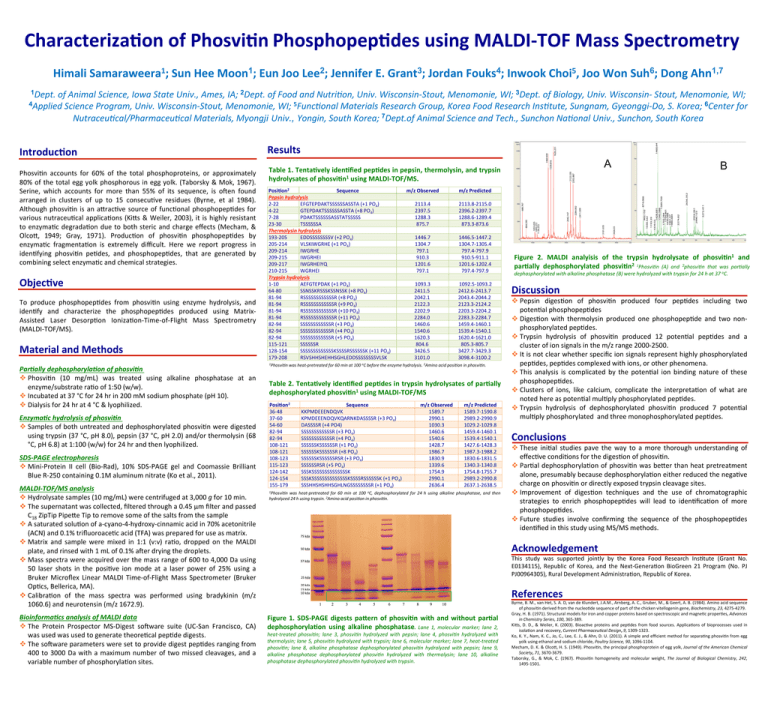
Characteriza*on of Phosvi*n Phosphopep*des using MALDI-­‐TOF Mass Spectrometry 1 1 2 3 4 5 6 1,7 Himali Samaraweera ; Sun Hee Moon ; Eun Joo Lee ; Jennifer E. Grant ; Jordan Fouks ; Inwook Choi , Joo Won Suh ; Dong Ahn 1Dept. of Animal Science, Iowa State Univ., Ames, IA; 2Dept. of Food and Nutri=on, Univ. Wisconsin-­‐Stout, Menomonie, WI; 3Dept. of Biology, Univ. Wisconsin-­‐ Stout, Menomonie, WI; 4Applied Science Program, Univ. Wisconsin-­‐Stout, Menomonie, WI; 5Func=onal Materials Research Group, Korea Food Research Ins=tute, Sungnam, Gyeonggi-­‐Do, S. Korea; 6Center for Nutraceu=cal/Pharmaceu=cal Materials, Myongji Univ., Yongin, South Korea; 7Dept.of Animal Science and Tech., Sunchon Na=onal Univ., Sunchon, South Korea Introduc*on Phosvi(n accounts for 60% of the total phosphoproteins, or approximately 80% of the total egg yolk phosphorous in egg yolk. (Taborsky & Mok, 1967). Serine, which accounts for more than 55% of its sequence, is oKen found arranged in clusters of up to 15 consecu(ve residues (Byrne, et al 1984). Although phosvi(n is an aPrac(ve source of func(onal phosphopep(des for various nutraceu(cal applica(ons (KiPs & Weiler, 2003), it is highly resistant to enzyma(c degrada(on due to both steric and charge effects (Mecham, & OlcoP, 1949; Gray, 1971). Produc(on of phosvi(n phosphopep(des by enzyma(c fragmenta(on is extremely difficult. Here we report progress in iden(fying phosvi(n pe(des, and phosphopep(des, that are generated by combining select enzyma(c and chemical strategies. Objec*ve To produce phosphopep(des from phosvi(n using enzyme hydrolysis, and iden(fy and characterize the phosphopep(des produced using Matrix-­‐ Assisted Laser Desorp(on Ioniza(on-­‐Time-­‐of-­‐Flight Mass Spectrometry (MALDI-­‐TOF/MS). Material and Methods Par%ally dephosphoryla%on of phosvi%n v Phosvi(n (10 mg/mL) was treated using alkaline phosphatase at an enzyme/substrate ra(o of 1:50 (w/w). v Incubated at 37 °C for 24 hr in 200 mM sodium phosphate (pH 10). v Dialysis for 24 hr at 4 °C & lyophilized. Enzyma%c hydrolysis of phosvi%n v Samples of both untreated and dephosphorylated phosvi(n were digested using trypsin (37 °C, pH 8.0), pepsin (37 °C, pH 2.0) and/or thermolysin (68 °C, pH 6.8) at 1:100 (w/w) for 24 hr and then lyophilized. SDS-­‐PAGE electrophoresis v Mini-­‐Protein II cell (Bio-­‐Rad), 10% SDS-­‐PAGE gel and Coomassie Brilliant Blue R-­‐250 containing 0.1M aluminum nitrate (Ko et al., 2011). MALDI-­‐TOF/MS analysis v Hydrolysate samples (10 mg/mL) were centrifuged at 3,000 g for 10 min. v The supernatant was collected, filtered through a 0.45 µm filter and passed C18 ZipTip PipePe Tip to remove some of the salts from the sample v A saturated solu(on of a-­‐cyano-­‐4-­‐hydroxy-­‐cinnamic acid in 70% acetonitrile (ACN) and 0.1% trifluoroace(c acid (TFA) was prepared for use as matrix. v Matrix and sample were mixed in 1:1 (v:v) ra(o, dropped on the MALDI plate, and rinsed with 1 mL of 0.1% aKer drying the droplets. v Mass spectra were acquired over the mass range of 600 to 4,000 Da using 50 laser shots in the posi(ve ion mode at a laser power of 25% using a Bruker Microflex Linear MALDI Time-­‐of-­‐Flight Mass Spectrometer (Bruker Op(cs, Bellerica, MA). v Calibra(on of the mass spectra was performed using bradykinin (m/z 1060.6) and neurotensin (m/z 1672.9). Bioinforma%cs analysis of MALDI data v The Protein Prospector MS-­‐Digest soKware suite (UC-­‐San Francisco, CA) was used was used to generate theore(cal pep(de digests. v The soKware parameters were set to provide digest pep(des ranging from 400 to 3000 Da with a maximum number of two missed cleavages, and a variable number of phosphoryla(on sites. Results A Table 1. Tenta*vely iden*fied pep*des in pepsin, thermolysin, and trypsin hydrolysates of phosvi*n1 using MALDI-­‐TOF/MS. Posi*on2 Sequence Pepsin hydrolysis 2-­‐22 EFGTEPDAKTSSSSSSASSTA (+1 PO4) 4-­‐22 GTEPDAKTSSSSSSASSTA (+8 PO4) 7-­‐28 PDAKTSSSSSSASSTATSSSSS 23-­‐30 TSSSSSSA Thermolysin hydrolysis 193-­‐205 EDDSSSSSSSSV (+2 PO4) 205-­‐214 VLSKIWGRHE (+1 PO4) 209-­‐214 IWGRHE 209-­‐215 IWGRHEI 209-­‐217 IWGRHEIYQ 210-­‐215 WGRHEI Trypsin hydrolysis 1-­‐10 AEFGTEPDAK (+1 PO4) 64-­‐80 SSNSSKRSSSKSSNSSK (+8 PO4) 81-­‐94 RSSSSSSSSSSSSR (+8 PO4) 81-­‐94 RSSSSSSSSSSSSR (+9 PO4) 81-­‐94 RSSSSSSSSSSSSR (+10 PO4) 81-­‐94 RSSSSSSSSSSSSR (+11 PO4) 82-­‐94 SSSSSSSSSSSSR (+3 PO4) 82-­‐94 SSSSSSSSSSSSR (+4 PO4) 82-­‐94 SSSSSSSSSSSSR (+5 PO4) 115-­‐121 SSSSSSR 128-­‐154 SSSSSSSSSSSSSKSSSSRSSSSSSK (+11 PO4) 179-­‐208 RSVSHHSHEHHSGHLEDDSSSSSSSSVLSK m/z Observed m/z Predicted 2113.4 2397.5 1288.3 875.7 2113.8-­‐2115.0 2396.2-­‐2397.7 1288.6-­‐1289.4 873.3-­‐873.6 1446.7 1304.7 797.1 910.3 1201.6 797.1 1446.5-­‐1447.2 1304.7-­‐1305.4 797.4-­‐797.9 910.5-­‐911.1 1201.6-­‐1202.4 797.4-­‐797.9 1093.3 2411.5 2042.1 2122.3 2202.9 2284.0 1460.6 1540.6 1620.3 804.6 3426.5 3101.0 1092.5-­‐1093.2 2412.6-­‐2413.7 2043.4-­‐2044.2 2123.3-­‐2124.2 2203.3-­‐2204.2 2283.3-­‐2284.7 1459.4-­‐1460.1 1539.4-­‐1540.1 1620.4-­‐1621.0 805.3-­‐805.7 3427.7-­‐3429.3 3098.4-­‐3100.2 1Phosvi=n was heat-­‐pretreated for 60 min at 100 oC before the enzyme hydrolysis. 2Amino acid posi=on in phosvi=n. Table 2. Tenta*vely iden*fied pep*des in trypsin hydrolysates of par*ally dephosphorylated phosvi*n1 using MALDI-­‐TOF/MS Posi*on2 36-­‐48 37-­‐60 54-­‐60 82-­‐94 82-­‐94 108-­‐121 108-­‐121 108-­‐123 115-­‐123 124-­‐142 124-­‐154 155-­‐179 Sequence KKPMDEEENDQVK KPMDEEENDQVKQARNKDASSSSR (+3 PO4) DASSSSR (+4 PO4) SSSSSSSSSSSSR (+3 PO4) SSSSSSSSSSSSR (+4 PO4) SSSSSSKSSSSSSR (+1 PO4) SSSSSSKSSSSSSR (+8 PO4) SSSSSSKSSSSSSRSR (+3 PO4) SSSSSSRSR (+5 PO4) SSSKSSSSSSSSSSSSSSK SSSKSSSSSSSSSSSSSSKSSSSRSSSSSSK (+1 PO4) SSSHHSHSHHSGHLNGSSSSSSSSR (+1 PO4) m/z Observed 1589.7 2990.1 1030.3 1460.6 1540.6 1428.7 1986.7 1830.9 1339.6 1754.9 2990.1 2636.4 m/z Predicted 1589.7-­‐1590.8 2989.2-­‐2990.9 1029.2-­‐1029.8 1459.4-­‐1460.1 1539.4-­‐1540.1 1427.6-­‐1428.3 1987.3-­‐1988.2 1830.6-­‐1831.5 1340.3-­‐1340.8 1754.8-­‐1755.7 2989.2-­‐2990.8 2637.1-­‐2638.5 1Phosvi=n was heat-­‐pretreated for 60 min at 100 oC, dephosphorylated for 24 h using alkaline phosphatase, and then hydrolyzed 24 h using trypsin. 2Amino acid posi=on in phosvi=n. (a) (b) B Figure 2. MALDI analyisis of the trypsin hydrolysate of phosvi*n1 and par*ally dephosphorylated phosvi*n2 1Phosvi=n (A) and 2phosvi=n that was par=ally dephosphorylated with alkaline phosphatase (B) were hydrolyzed with trypsin for 24 h at 37 oC. Discussion v Pepsin diges(on of phosvi(n produced four pep(des including two poten(al phosphopep(des v Diges(on with thermolysin produced one phosphopep(de and two non-­‐ phosphorylated pep(des. v Trypsin hydrolysis of phosvi(n produced 12 poten(al pep(des and a cluster of ion signals in the m/z range 2000-­‐2500. v It is not clear whether specific ion signals represent highly phosphorylated pep(des, pep(des complexed with ions, or other phenomena. v This analysis is complicated by the poten(al ion binding nature of these phosphopep(des. v Clusters of ions, like calcium, complicate the interpreta(on of what are noted here as poten(al mul(ply phosphorylated pep(des. v Trypsin hydrolysis of dephosphorylated phosvi(n produced 7 poten(al mul(ply phosphorylated and three monophosphorylated pep(des. Conclusions v These ini(al studies pave the way to a more thorough understanding of effec(ve condi(ons for the diges(on of phosvi(n. v Par(al dephosphoryla(on of phosvi(n was bePer than heat pretreatment alone, presumably because dephosphoryla(on either reduced the nega(ve charge on phosvi(n or directly exposed trypsin cleavage sites. v Improvement of diges(on techniques and the use of chromatographic strategies to enrich phosphopep(des will lead to iden(fica(on of more phosphopep(des. v Future studies involve confirming the sequence of the phosphopep(des iden(fied in this study using MS/MS methods. Acknowledgement This study was supported jointly by the Korea Food Research Ins(tute (Grant No. E0134115), Republic of Korea, and the Next-­‐Genera(on BioGreen 21 Program (No. PJ PJ00964305), Rural Development Administra(on, Republic of Korea. References Figure 1. SDS-­‐PAGE digests paVern of phosvi*n with and without par*al dephosphoryla*on using alkaline phosphatase. Lane 1, molecular marker; lane 2, heat-­‐treated phosvi=n; lane 3, phosvi=n hydrolyzed with pepsin; lane 4, phosvi=n hydrolyzed with thermolysin; lane 5, phosvi=n hydrolyzed with trypsin; lane 6, molecular marker; lane 7, heat-­‐treated phosvi=n; lane 8, alkaline phosphatase dephosphorylated phosvi=n hydrolyzed with pepsin; lane 9, alkaline phosphatase dephosphorylated phosvi=n hydrolyzed with thermolysin; lane 10, alkaline phosphatase dephosphorylated phosvi=n hydrolyzed with trypsin. Byrne, B. M., van Het, S. A. D, van de Klundert, J.A.M., Arnberg, A. C., Gruber, M., & Geert, A. B. (1984). Amino acid sequence of phosvi(n derived from the nucleo(de sequence of part of the chicken vitellogenin gene, Biochemistry, 23, 4275-­‐4279. Gray, H. B. (1971). Structural models for iron and copper proteins based on spectroscopic and magne(c proper(es, Advances in Chemistry Series, 100, 365-­‐389. KiPs, D. D., & Weiler, K. (2003). Bioac(ve proteins and pep(des from food sources. Applica(ons of bioprocesses used in isola(on and recovery, Current Pharmaceu=cal Design, 9, 1309-­‐1323. Ko, K. Y., Nam, K. C., Jo, C., Lee, E. J., & Ahn, D. U. (2011). A simple and efficient method for separa(ng phosvi(n from egg yolk using ethanol and sodium chloride, Poultry Science, 90, 1096-­‐1104. Mecham, D. K. & OlcoP, H. S. (1949). Phosvi(n, the principal phosphoprotein of egg yolk, Journal of the American Chemical Society, 71, 3670-­‐3679. Taborsky, G., & Mok, C. (1967). Phosvi(n homogeneity and molecular weight, The Journal of Biological Chemistry, 242, 1495-­‐1501.


