elps5092-sup-0004-Suppmat
advertisement
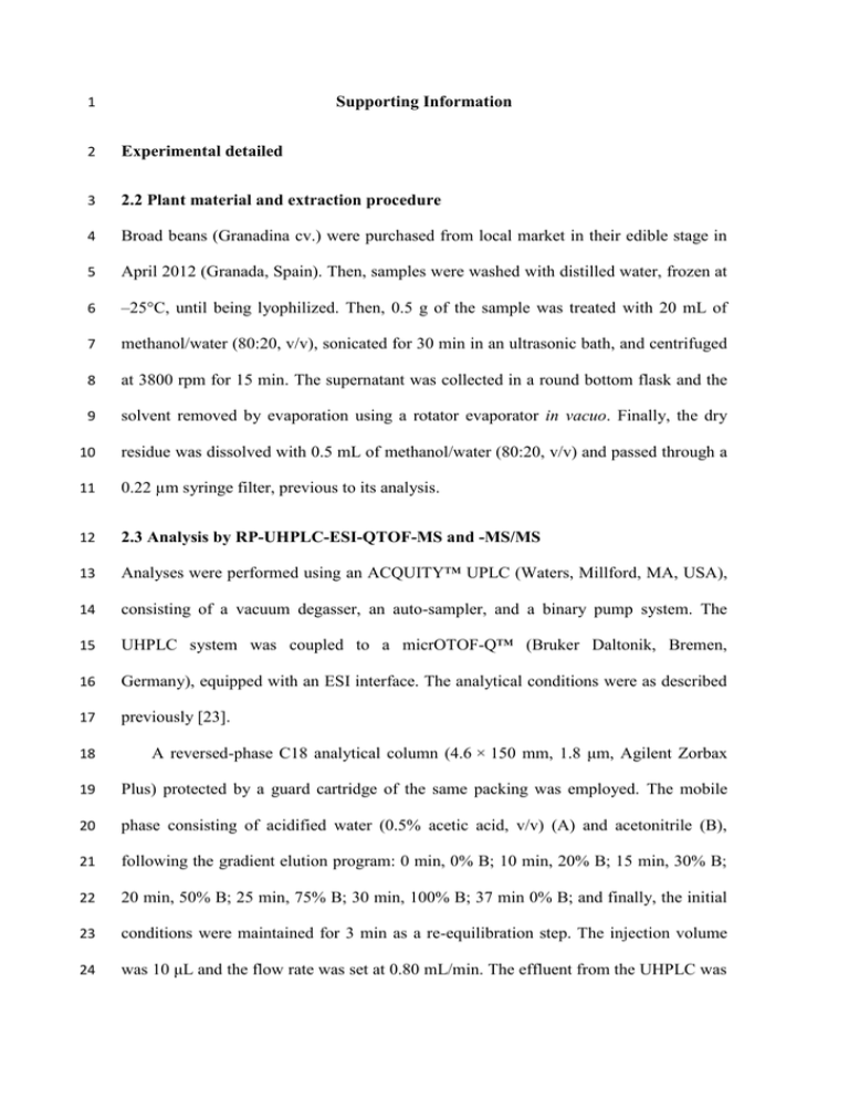
1 Supporting Information 2 Experimental detailed 3 2.2 Plant material and extraction procedure 4 Broad beans (Granadina cv.) were purchased from local market in their edible stage in 5 April 2012 (Granada, Spain). Then, samples were washed with distilled water, frozen at 6 –25°C, until being lyophilized. Then, 0.5 g of the sample was treated with 20 mL of 7 methanol/water (80:20, v/v), sonicated for 30 min in an ultrasonic bath, and centrifuged 8 at 3800 rpm for 15 min. The supernatant was collected in a round bottom flask and the 9 solvent removed by evaporation using a rotator evaporator in vacuo. Finally, the dry 10 residue was dissolved with 0.5 mL of methanol/water (80:20, v/v) and passed through a 11 0.22 µm syringe filter, previous to its analysis. 12 2.3 Analysis by RP-UHPLC-ESI-QTOF-MS and -MS/MS 13 Analyses were performed using an ACQUITY™ UPLC (Waters, Millford, MA, USA), 14 consisting of a vacuum degasser, an auto-sampler, and a binary pump system. The 15 UHPLC system was coupled to a micrOTOF-Q™ (Bruker Daltonik, Bremen, 16 Germany), equipped with an ESI interface. The analytical conditions were as described 17 previously [23]. 18 A reversed-phase C18 analytical column (4.6 × 150 mm, 1.8 μm, Agilent Zorbax 19 Plus) protected by a guard cartridge of the same packing was employed. The mobile 20 phase consisting of acidified water (0.5% acetic acid, v/v) (A) and acetonitrile (B), 21 following the gradient elution program: 0 min, 0% B; 10 min, 20% B; 15 min, 30% B; 22 20 min, 50% B; 25 min, 75% B; 30 min, 100% B; 37 min 0% B; and finally, the initial 23 conditions were maintained for 3 min as a re-equilibration step. The injection volume 24 was 10 μL and the flow rate was set at 0.80 mL/min. The effluent from the UHPLC was 25 splited using a T-type splitter and directed to the mass spectrometer at approximately 26 0.2 mL/min. The spectra were acquired in negative-ion mode over a mass-to-charge 27 (m/z) range from 50 to 1100. QTOF parameters were set at: capillary voltage, +4.0 kV; 28 drying gas temperature, 210°C; drying gas flow, 8.0 L/min; nubilizing gas pressure, 29 2 bar; end-plate offset, –0.5 kV. Moreover, automatic MS/MS was set. Nitrogen was 30 used as drying, nebulising and collision gas. A sodium acetate cluster solution was used 31 for external instrument calibration. 32 The MS and MS/MS data were processed through Data Analysis 4.0 software 33 (Bruker 34 (SmartFormula), simulate isotope patterns, as well as theoretical monoisotopic, 35 nominal, and average masses (IsotopePattern), and build common neutral losses 36 (BuildingBlockEditor). Therefore, the elemental composition of each compound was 37 determined by the SmartFormula tool that generates a list of possible molecular 38 formulae based on a CHNO algorithm, and the other elements which usually occur in 39 smaller numbers could be also added. It provides standard functionalities such as 40 minimum/maximum elemental range, electron configuration, and ring-plus double-bond 41 equivalents, the deviation between the measured mass and theoretical mass (error) (Da 42 and ppm), and a sophisticated comparison of the theoretical with the measured isotope 43 patterns (msigma value or m) for increasing the confidence in the suggested molecular 44 formula. The broadly accepted error and m value for confirmation of elemental 45 compositions were set at 5 ppm and at 50, respectively. The use of isotopic-abundance 46 pattern removes more than 95% of false candidates. The molecular formula generated 47 for each compound, its MS/MS spectra and its comparison with spectra found in the 48 literature was the main tool for putative identification of broad bean metabolites. For 49 this, the following chemical structure databases were consulted: PubChem Daltonics), which supplies tools to calculate molecular formulae 50 (http://pubchem.ncbi.nlm.nih.gov), 51 SciFinder Scholar (https://scifinder.cas.org), HMDB (http://www.hmdb.ca/), Phenol- 52 Explorer 53 (http://kanaya.naist.jp/knapsack_jsp/top.html), Metlin (http://metlin.scripps.edu), and 54 MassBank (http://www.massbank.jp), which is related to KNApSAcK database. The 55 two latter databases, Metlin and MassBank, also include a repository of tandem mass 56 spectrometry data. 57 ChemSpider (www.phenol-explorer.eu), (http://www.chemspider.com), KNApSAcK Core System 58 Results detailed 59 3.4 Structural prediction of unknown metabolites from V. faba (detailed) 60 Along the consciously profiling of the seed extract, 24 compounds did not match 61 adequately with those found in literature and databases sharing the same molecular 62 formula. Thus, modifications of the previous characterized metabolites through 63 common metabolic reactions [72] were established according to their MS and MS 2 data: 64 hydroxylation (+ O), methylation (+ CH2), conjugation with leucine/isoleucine (+ 65 C6H11NO2), and glycosylation (with hexose, C6H10O5, and with pentose, + C5H8O4). 66 These added groups could be assigned directly as fragment ions or indirectly as neutral 67 losses in MS2. The compounds proposed were derivatives of vicine, convicine, eucomic 68 acid, jasmonic acid, and adenosine. To make easy the comprehension, the fragments 69 shared in common or related to those are showed in bold letter in Table S2 (Supporting 70 Information). 71 Several isomers of vicine and convicine conjugated with hexose (compounds A-E) 72 and pentose (compounds F-I) were found. In MS2 the sequential losses of the sugars 73 leaded to the aglycones divicine and isouramil, and, afterwards, the loss of NHO to m/z 74 110.0341 and 111.0188 (uracil), respectively. In the case of convicine derivatives, the 75 fragment ion at m/z 141.0243 corresponded to the odd ion of isouramil. Moreover, two 76 consecutive losses of CHNO from pyrimidone ring of convicine derivatives were also 77 observed, in agreement with results commented above. On the other hand, the purine 78 nucleoside derivative succinyladenosine (J) was tentatively identified according to the 79 MS and MS/MS data. In this case, the fragmentation pattern was similar to that 80 described above for adenosine and correlated well with our proposal. Thus, the main 81 fragment ions were at m/z 266.0928, 250.0591 and 206.0695, corresponding to the loss 82 of the succinic acid moiety, the ribose moiety and the successive decarboxylation, 83 whereas m/z 134.0464 and m/z 115.0033 were adenine and the succinic acid moiety 84 ([C4H6O4 – H2 – H]-), respectively. This compound is a biomarker of purine metabolism 85 disorders in humans [78], so the potential presence in edible vegetables deserves further 86 studies. 87 The other putative metabolites were related to jasmonic acid and dihydrojasmonic 88 acid. In the MS2 spectra of the hydroxylated and glycosylated forms (compounds K-M 89 and P), subsequent neutral losses of H2O according to the number of the OH groups, 90 and/or the hexose moiety was observed, together with the loss of CO2 from the carboxyl 91 group of the jasmonate backbone. In that case, fragment ions at m/z 179.1073 92 ([C11H16O2 – H]-) from compounds K, L and P and m/z 177.0928 ([C11H14O2 – H]-) 93 from compound M resemble fragment ion m/z 181.1224 ([C11H18O2 – H]-) found in 94 MS2 spectrum of compound OH-JA-Ile (see Fig. 2C). On the other hand, 95 leucine/isoleucine was detected everywhere in compounds S-X and the product ion at 96 m/z 142.0874 ([C7H13NO2 – H]-). Alternatively, compounds O and Q gave a major 97 fragment ion at m/z 146.0812 with molecular formula C6H13NO3, indicating that could 98 be hydroxyleucine. Regarding to this, 4-hydroxyisoleucine has been described in 99 Fabaceae [79]. 100 Two derivatives of eucomic acid were also proposed as methyl eucomic acid 101 (compound N, m/z 269.0675) and benzylmalic acid (compound R, m/z 223.0612). 102 According to the fragmentation pattern of eucomic acid, the fragmentation mainly 103 occurred at the malic acid moiety. Thus, compounds N and R showed the major product 104 ions at m/z 209.0462 and 163.0399, respectively, corresponding to the loss of 105 CH3−COOH (60.0211 u). Moreover, the loss of H2O and CO2 at m/z 207.0666 and 106 161.0601, together with successive losses of C2H4 at m/z 179.0718 and m/z 133.0648, or 107 CO2 at m/z 163.0745 and m/z 117.0702, respectively, reinforce the assignments. In the 108 case of the compound N, the loss of malic acid – H2 (C4H4O5, 132.0059 u) was clearly 109 observed at m/z 137.0603, and subsequently the loss of CH3 at m/z 122.0381. 110 111 Other references 112 [78] Ito, T., van Kuilenburg, A. B.P., Bootsma, A. H., Haasnoot, A. J., van Cruchten, 113 A., Wada, Y., van Gennip, A. H., Clin. Chem. 2000, 46, 445–452. 114 [79] Jetté, L., Harvey, L., Eugeni, K., Levens, N., Curr. Opin. Investig. Drugs 2009, 10, 115 353-358.

