Examples
advertisement
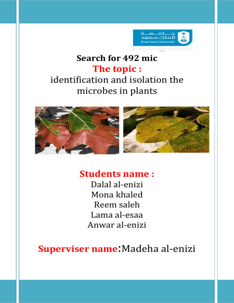
Search for 492 mic The topic : identification and isolation the microbes in plants Students name : Dalal al-enizi Mona khaled Reem saleh Lama al-esaa Anwar al-enizi Superviser name:Madeha al-enizi Contents: Introduction……………………………..………………………...... Pathogene microbes in plant ………….. Benefite microbes in plant……………. Materials and methods………………………………………… a.Virus………………………..………………………………………… b.Bacteria…………………….……………………………………….. c. Fungi……………………….………………………………………... Results............................…….……...…………………………... Discussion……………………………………….………. Recommendations………………………………………………… References………………………..…………………………………… Identification and Isolation the microbes (Virus, Bacteria Fungi,) in plants Introduction: Pathogenemicrobes in plants : Every gardener has put in plants with hopes for wonderful flowers, fruits, or vegetables, only to have those hopes dashed as the plants get sick and die. Plants that die are considered diseased. Many things can cause plants to become diseased, including living agents, other factors (nonliving), or a combination of the two. This chapter focuses only on living agents— fungi, bacteria, viruses, nematodes, and parasitic plants. Nonliving factors, such as nutrient deficiencies, lack of water, temperature stress, and these problems in combination as they relate to specific types of plants, are discussed elsewhere. Adapted for Kentucky by John Hartman, extension plant pathologist, University of Kentucky. Viruses: Virus particles are composed of a few strands of molecules and are even smaller than bacteria. Even with a high-magnification microscope, they can be seen only when massed together in a plant cell. Electron microscopes reveal them to have many shapes, including long strands, short rods, or multi-sided balls. Viruses use a host plant’s cell organelles (cell organs) to produce more viruses. The result can be strange plant colors, forms, or structures. Some viral infections, however, don’t result in any visible plant problems. Touching virus-infected plant material and then touching healthy plants can transmit some viruses. For example, a smoker can transmit tobacco mosaic virus from a cigarette to a plant. Many other viruses are transmitted by insects such as aphids, scales, or whiteflies. Fungi, mites, nematodes, and even parasitic plants also can transmit viruses. Viruses also may infect a host plant’s seeds and thus be transmitted to the next generation. Examples: 1- Beet curly top virus sp. 2- Alfalfa mosaic virus sp. Fungi In general, they are multicellular organisms with a threadlike body. These filamentlike threads, which are called hypha, have cell walls. When many threads mass together, they form what is called a mycelium, a mass of interwoven threads. Further growth of a mycelium may produce fruiting bodies, in which sexual or asexual spores are formed. The mycelium, fruiting bodies, and spores are used to identify and diagnose fungal problems. Some fungi can survive and grow without a living host. Others die if they are not closely associated with a host. Fungi cause plant diseases in one of the following ways: • making toxins that kill plant cells. • growing within a plant’s vascular system and plugging it up . • rotting roots (which cuts off the leaves’ supply of water and nutrients). • sending rootlike structures into plant cells. Examples: 1- Pythium sp. 2- Phytophthora sp. Bacteria: Bacteria are single-celled organisms that are much smaller and less complex than plant cells. Many are about the size of a plant chloroplast. Their cell walls are covered with a slimy material known as a “matrix,” and they reproduce by splitting in two. Bacteria can build up to such high numbers in a plant that they ooze out the plant’s tissues and may attract insects that spread the bacteria to healthy plants.. Bacteria cause plant diseases by forming toxins or by producing enzymes that break down plants’ cell walls. Crown gall bacteria actually genetically engineer their host plant to make galls and amino acids, thus giving the bacteria a place to live (the galls) and the chemicals they need to grow and reproduce (the amino acids). Examples: 1- (Pseudomonas syringaepv. tomato) 2- (Corynebacterium michiganensis) 3- (Psuedomonas corrugata) 4- (Ralstonia solanacearum) 5- (Xanthomonas euvesicatoria) o Disease is a result of simultaneous interactions between the environment, host, and pathogen. Disease Cycle: The sequence of events from a pathogen’s survival to plant disease development and back to pathogen survival is called the disease cycle, or the pathogen’s life history. By understanding of the disease cycle— learning the chain of events that contribute to a disease—you can find the weakest links and take measures to break the cycle. Most pathogens must survive a period of adverse conditions, usually winter, when they do not actively cause disease. The host plant is infected or continues to be infected by the pathogen’s overwintered disease-transmitting substance, inoculum, in the spring. Some diseases, such as many canker diseases, have only one cycle during the year. Others, such as powdery mildew, continually produce more inoculum, thus repeating the cycle many times during a single growing season. Disease Diagnosis There is no single set of questions or technique for diagnosing plant diseases. Experience and practice are the best teachers. Plant problems are most easily diagnosed through personal, on-site inspection. You may notice subtle influences of the site, environment, or management practices that make identification easier. Diagnosis is more difficult when only part of a plant is seen, which may or may not indicate the real problem. The worst diagnostic method is by phone, which easily can lead to misunderstanding and inaccuracy. Conditions Necessary for Pathogenic Diseases In order for a pathogenic (biotic) plant disease to occur, three conditions must bemet: • The host plant must be susceptible. • An active, living pathogen must be present. • The environment must be suitable or favorable for disease development. Benefite microbes in plants : bacteria : New research suggests that microbes could provide an alternative to existing agricultural methods and genetic engineering in alleviating some of these problems. For instance, sunflowers and some other plants naturally produce the sugar trehalose, which helps to stabilize plant cell membranes and to reduce the damage from cycles of drying followed by rehydration. Stores carbohydrate Protects cells against various stress conditions Other plants, including corn and potatoes, have been genetically engineered to manufacture trehalose. Yet molecular biologist (Gabriel ,2013)in Mexico hopes to eventually treat crops without any genetic modification by using the trehalose-producing bacterium Rhizobium etli, which is found around the roots of bean plants. Also make thenitrogene fixation . An earlier experiment with a genetically altered version of the bacterium improved yields by 50 percent in normal conditions—and saved half the crop during a drought. Fungi : Mycorrhizaearesymbiotic relationships that form between fungi and plants. The fungi colonize the root system of a host plant, providing increased water and nutrient absorption capabilities while the plant provides the fungus with carbohydrates formed from photosynthesis. Mycorrhizae also offer the host plant increased protection against certain pathogens. What Benefits do Mycorrhizal Fungi Offer to Plants? Fungi are heterotropic organisms, and must absorb their food. Fungi also have the ability to easily absorb elements such a phosphorus and nitrogen which are essential for life. Plants are autotropic, producing their food in the form of carbohydrates through the process of photosynthesis. However, plants often have difficulty obtaining and absorbing many of the essential nutrients needed for life, specifically nitrogen and phosphorus. © 2003 The New York Botanical Garden Ectomycorrizae Endomycorrizae Copyright 2010 Survivallandusa.com Materials and methods: A. Virus: 1-Peanut bud necrosis virus Virus isolates: The Peanut bud necrosis virus suspecting onion plants (n=145) were collected from different places inIndia. The PBNV infected onion plants showed symptoms like straw colored, mosaic and necrotic lesions on the young leaves of onion stalks (Fig.1). 2- Enzyme linked immunosorbent assay (ELISA): The onion samples were collected from the different places in South India ELISA (DAC-ELISA) [15] using -subjected to direct antigen coating specific polyclonal TSV antiserum. The suspected leaf samples were groundin carbonate buffer (pH 9.6) at 1:10 dilution (w/v) and crude leaf extracts were used as antigens. The healthyleaf tissue extract was used as a control. 100μl of each sample extract was loaded into wells of ELISA plates(NuncMaxiSorb; Denmark) and incubated at 37°C for 60 min. The antigen coated plates were washed threetimes with PBS-T buffer. The plates were then blocked with blocking buffer (PBS-TPO with 5% skimmed milkpowder) and incubated at 37°C for another 60 min. The plates were then washed with PBS-T as described above. The polyclonal antisera of PBNV was used as primary antibodies at 1:5000 (v/v) dilutions in antibody buffer (PBS-TPO- 0.15 M NaCl in 0.1 M phosphate buffer pH 7.4, 0.05% Tween 20, 2% polyvinyl pyrrolidine, 0.2% ovalbumin), incubated for 60 min at 37°C and washed three times with PBS-T as above. Goat antirabbit- (A. Sujitha et al.2012) ALP conjugate (Sigma, Germany) at 1:10000 (v/v) dilution in antibody buffer was added and incubated at 37°Cfor 60 min. Para-nitrophenyl phosphate (PNPP) (Sigma, Germany) was used as a substrate at 5 mg / 10 ml ofsubstrate buffer (Diethanolamine buffer, pH 9.8). Absorbance values were recorded in an ELISA plate reader(Bio-Rad, USA) at 405 nm after 15-30 min of substrate addition. Antigen buffer control was included alongwith leaf antigen samples. The reactions were terminated using 3 N NaOH (50μl/well). Only positive sampleswhose A405 values were twice or more greater than twice the value of healthy onion samples was considered asvirus infected. The ELISA positive samples were maintained in local lesion assay host as Vignaunguiculata(cv-C 152)cultivar was used following sub-culturing for three generations on the same host and later maintained on theirnatural hosts for further studies. Preparation of total RNA and RT-PCR Total RNA from 100 mg of healthy and GBNV infected onion leaf samples was isolated using RNeasyPlantMinikit according to the manufacturer’s instructions (Qiagen, Germantown, MD, USA). The resulting totalRNA was incubated with GBNV-CP gene-specific reverse primer at 65°C for 5 min and snap-chilled on ice for2 min. cDNA was synthesized using M-MuLV reverse transcriptase (Fermentas, Ontario, Canada) at 42°C for 1 h. The genome sense primer, 5′ATGTCTAACGT(C/T) AAGCA (A/G) CTC 3′, and antisense primer,5′TTACAATTCCAGCGAAGGACC 3, were used to amplify the complete CP gene of GBNV [16]. Twomicrcolitre of cDNA was amplified in a 25 μl reaction volume containing 2.5 U of Taq DNA Polymerase(Fermentas), 10 pmol of forward (GBNV-CP-F) and reverse primer (GBNV-CP-R), 2.5 mM MgCl2 and 10 mMeachdNTP’s. PCR amplification conditions included an initial denaturation cycle of 5 min at 94°C, followed by35 cycles of denaturation for 30 s at 94°C, annealing for 1 min at 56°C and extension for 1 min at 72°C withfinal extension for 60 min at 72°C. Amplified products were resolved following electrophoresis through a 1%agarose gel containing ethidium bromide (10 mg/ml). B. Bacteria: 1-Isolation of bacteria The infected leaves were collected from S146 and TR10 mulberry varieties based on various symptoms of leaf blight between January 2010 and December 2010. The infected leaves were brought to the laboratory and the bacterial pathogen was isolated by following the microbial culture method suggested by Sulle and Seemuller (1987). The portion of infected leaf was cut into small pieces and sterilized by dipping them in 70% ethanol for 10 seconds and then in 2% sodium hypochlorite for 2 min. The leaf samples were washed thoroughly in sterile distilled water for three times and macerated in sterile water (1 g/ml), kept for 10 min to obtain a suspension and 1 ml of suspension was inoculated under sterile conditions. The culture medium consists of King’s medium B /nutrient agar mixed with 5 mg/l antifungal agent (Griseofulvin). Spread plate technique was used for inoculation. The culture plates were incubated at 26-28˚C for 3-5 days. Optical characterization of bacterial suspensions was carried out using UV spectrophotometer (Thermo) at OD600 = 0.265 which corresponds to a cell density of 1x10⁸ CFU/ml. Single colonies were sub-cultured on King’s medium-B by following methods suggested by King et al. (1954). 2-Staining procedures The colonies were stained using simple stain, gram stain, spore stain and capsule stain for examination of shape and arrangement of bacterial cells. The bacterial colonies were distinguished into groups viz. gram-positive or gram-negative. Different biochemical tests such as KOH test, fluorescent pigmentation test, carbohydrate fermentation test, gelatin liquefaction test, catalase test, starch hydrolysis test, asein hydrolysis test, oxidase test, litmus milk test, indole production, M–R test, V–P test, citrate utilization test and hydrogen sulphide production test were performed. Fig. 1. The scanning electron micrograph of Pseudomonas syringae 3-Sample preparation for Scanning Electron Microscope (SEM) Bacterial isolates were harvested from culture plates and fixed with few drops of 2.5% glutaraldehyde for 2-4 hrs at 4°C, washed using 0.1 M phosphate buffer for 3 times, centrifuged at 3000 to 5000 rpm each of 15 min at 4°C to collect pellet and the supernatant was discarded. 1% osmium tetroxide was used as post fixative for 2 hrs at 4°C and the bacterial suspension was washed in 0.1 M phosphate buffer for 3 times each of 15 min at 4°C to remove the unreacted fixative. The bacterial suspension was mounted on the aluminium stubs using carbon adhesive tape and frozen using liquid nitrogen. The samples were viewed using a scanning electron microscope (JEOL 6490 LV) by applying different accelerating voltage (Stadtlander et al., 2007) C. Fungi: 1- Fungal isolate Penicillium notatum was isolated from rhizosphere soil of planted Chinese mustard (Brassica campestrisvar. chinensis) using the dilution plate technique on Czapek's agar (2g sodium nitrate, Ig potassium dibasic phosphate, 0/5g potassium chloride, 0.5g magnesium sulphate, l7g agar and 1,000 ml of distilled water). 2-Testing for plant growth: Penicillium notatum(KMITL 99) was cultured on potato dextrose agar (PDA) and incubated for 14 days at room temperature (27-30 C). Medium was sterilizeu al 121 C for 20 minutes for 3 consecutive days and placed into 12 inches diameter plastic pots. Fourteen days old P. notatumplates were harvested and added to the planting medium at concentrations of 5.53 x 10⁶, 11.06 x 10⁶, 16.59 x 10⁶, 22.13 x 10⁶ and 27.66 x 10⁶ spores/ml at a rate of 50 ml/pot. Planting medium without the addition of P. notatum was used as a control. All pots were moistened with sterile water and covered with plastic sheets and left for 15 days before planting. The effect of P. notatumon growth of the Chinese mustard, Chinese radish and cucumber were carried out. Ten seeds of each plant were sown in each pot and the plants were thinned randomly to 1 seedling per pot after emergence. Chemical fertilizers or pesticides were not applied in these experiments. Statistical analyses3-: At harvesting, the plant height, root length, root diameter, fresh and dry weights of the root and shoots were measured at 57 days for the Chinese mustard, 58 days for the cucumber and 73 days for the Chinese radish. The average mean of growth parameters from five replicated experiments were subjected to analysis of variance and treatment. Results: Fig. 1: Symptoms associated with natural occurrence of Peanut bud necrosis virus on onion: straw colored, mosaic and necrotic lesions were observed on the young leaves. ELISA: Table 1.Collection and screening of Peanut bud necrosis virus (PBNV) infecting onion samples fromdifferent locations in South India. Discussion: Feg1 and table 1:ThePBNV has a wide host range and it infects several hosts such as cowpea, groundnut, mung bean, potato (Bhat AI et al.2001) (Thien HX et al.2003) (Jain RK et al.2004). This work presents the sensitivitycomparison of two diagnostic methods, RT-PCR and ELISA in the detection of PBNV in onion. The symptomsof GBNV first appeared in the young leaflets such as straw colored, mosaic and necrotic lesions. If the PBNVinfection occurs at a young age, it results in the death of the plant due to severe necrosis. The PBNV infectedonion samples were confirmed by ELISA using the polyclonal antibodies of coat protein gene of PBNV. Theconfirmation of ELISA positive samples were maintained in cowpea (Cv-c-152). After five days sapinoculation, both localized and systemic infections were observed on cowpea plants. direct antigen coating enzyme linked immunosorbent assay (DACELISA) by using the PBNV specificantiserum, where as 124 samples (85.51%) were confirmed by RT-PCR. A single band of the expected size of~800 bp corresponding to the coat protein gene of PBNV was observed when total RNA extracted from infectedtissue was used in RT-PCR. (Table 2). Bacterial leaf blight was observed in the leaves of S146 and TR10 mulberry varieties during rainy season. The infected leaves were brown with irregular spots on the surface. These lesions were dark green at the beginning of infection, then turned yellow and eventually become brown. The infection spreads to the entire tree and leaves fall off prematurely. The colonies were shiny white and yellow, translucent, convex and circular with smooth surface and margin. The average colony diameter was 12 ± 2.45 mm. The isolate was gram negative rod, produced red or pink colour when counterstained with safranine. The biochemicaltests such as KOH test, fluorescent pigmentation test, carbohydrate fermentation test, gelatin liquefaction test, catalase test, casein hydrolysis test, litmus milk test and V-P test showed positive response . The results of present study clearly demonstrate that the leaf blight in mulberry leaves was caused by P. mori.. The biochemical tests such as KOH, fluorescent pigmentation, carbohydrate fermentation, catalase, casein hydrolysis, litmus milk, V-P tests showed positive response while gelatin liquefaction, starch hydrolysis, oxidase, indole production, M-R hydrogen sulphide production tests showed negative results. The results of biochemical tests in the present study confirmed that the observed pathogenic bacterium is P. mori. (Fig. 2): The plant height gradually increased when the concentration of P. notatumadded was increased from 5.53 x 10⁶ spores/ml to 16.59 x 10⁶ spores/m1. Further increases in concentrations of P.notatumto 22.13 x 10⁶ and 27.66 x 10⁶ spores/m1., however, resulted to gradual reduction of the plant height. Application of P. notatumat a concentration of 16.59 x 10⁶ spores/m1. resulted in plant growth that was significantly higher than the control (P = 0.05). Similar trends were observed in the other parameters measured but no significant differences were recorded. There was significant effect on growth of the Chinese radish when P. notatum was added. The Chinese radish shoot length, root length, total length. The results of these experiments reveal the potential of P. notatum(KMITL 99) for increasing plant growth, especially in the Chinese radish. Thetype of growth promotion may be similar to that produced by the addition ofTrichodermaspp. which have been found to enhance the growth of variousplants (Chang et al., 1986; Paulitzet al., 1986; Windham et al., 1986; Baker,1988; Kleifeld and Chet, 1992; Ousleyet al., 1994 a,b; Phuwiwat and Soy tong, 1999 a,b; Harman, 2000). The increased plant growth induced by Trichodermaspp., has been found to be dependent on many factors such as plants, the strain of Trichoderma used, the form of inoculum, the concentrations of inoculum applied, and the soil environment (Paulitzet al., 1986; Baker, 1988; Kleifeld and Chet, 1992; Ousleyet al., 1994a,b). It was study , P. notatum (KMITL 99) had no significant effect on the growth of Chinese mustard and no effect on the growth of cucumber plants. Some reports have shown that P. chrysogenum is able to control Verticillium wilt of tomato (Dutta, 1981). Interestingly, Mishra and Tiwan (1976) reported that P. notatum could inhibit and reduce the number of rust pustules in wheat caused by Puccinia graminis f. sp. tritici. It would be interesting to test this specific strain of P. notatum (KMITL 99) for biological control of plant pathogens. Recommendations: 1-We must be at the plant genes resistant microbe that injury may occur. 2-Soil must be used for the cultivation of the plant free of pathogens. 3-Plant must be done properly from pathogenic microbes and there is no help to the entry wounds. References: Plant Diseases ,Kentucky Master Gardener Manual Chapter 6 , By Jay W. Pscheidt, extension plant pathologist, Oregon State University. Edited by Lindsey du Toit, plant diagnostician, Washington State University-Puyallup, and Warren Copes, ornamental plant pathologist, Washington State University . Adapted for Kentucky by John Hartman, extension plant pathologist, University of Kentucky © 2003 The New York Botanical Garden. Baker, R. (1988). Trichodermaspp.as plant growth stimulants. CRC Critical Review of Biotechnology 7: 97-106. Kleifeld, O. and Chet, 1. (1992). Trichodermaharzianuminteraction with plants and effects on growth response. Plant and Soil 144: 267-272 . Kleifeld, O. and Chet, 1. (1992). Trichodermaharzianuminteraction with plants and effects on growth response. Plant and Soil 144: 267-272. Mishra, R.R. and Tiwari, R.P. (1976). Studies on biological control of Pucciniagraminis tritici. In: Microbiology of Aerial Plant Surfaces (eds. C.H. Dickinson and T.F. Preece). Academic Press, London Phuwiwat, W. and Soy tong, K. (1999a). Growth and yield response of Chinese radish to application of Trichodermaharzianum. Thammasat International Journal of Science andTechology 4: 68-71. Picci, G. (1965). Intornoallamicroflorapresentesulle olive sane e colpite da cicloconio. Agricultura Italia Pisa 65: 1-11. Dhar A, Bindroo BB (2000). Management of mulberry pests and diseases in north India. In: Seth MK, Agrawal HM, editors. Sericulture in India. 2: 457-475. King EO, Ward WK, Raney DE (1954). Two simple media for the demonstration of pyocyanin and fluorescein. J Laboratory & Clinical Medicine 44: 301-307. Narayanasamy P (2008). Molecular Variability of Microbial Plant Pathogens.Molecular Biology in Plant Pathogenesis and Disease Management. Microbial Plant Pathogens 1: 159-225. Ogra RK (2000). Diseases of mulberry and their control. In: Seth MK, Agrawal HM editors. Sericulture in India 2: 427-438. Pfleger FL, Gould SL (2009). Bacterial Leaf Diseases of Foliage Plants. University of Minnesota, College of Agriculture, Food and Environmental Science 5: 315-318. Purohit SS (2006).Nature of plant diseases. Microbiology Fundamentals and Applications 7: 667-672. Shree MP, Santakumari M (1994). Mulberry disease and their control. In: Boraiah G, editor. Lectures on sericulture; pp 19-23. Stadtlander CTK-H (2007). Scanning electron microscopy and transmission electron microscope of Mollicutes: Challenges and Opportunities. Morden research and educational, USA 3: 122-31. Sulle S, Seemuller E (1987). The role of ice formation in the infection of sour cherry leaves by Pseudomonas syringaepv. syringae. The American PhytopathSoc 77(2): 173-177. Tang KMA, Salam KA, Samad MA, Absar N (2005). Nutritional changes of four varieties mulberry leaves infected with fungus (Cercosporamoricola). Pak J BiolSci 8(1): 127-131. USDA Forest Service (2005).Forest Health Staff, Newton Square, A weed of the week; pp. 12-13. Yoshida S, Shirata A, Hiradate S (2002). Ecological characteristics and biological control of mulberry anthracnose.JARQ 36(2): 89-95. Young JM, Dye DW, Bradbury JF, Panagopoulos CG, Robbs CF (1978). A proposed nomenclature and classification for plant pathogenic bacteria. NZJ Agric Res 21: 153-177. Jones, J.B., Jones, J.P., Stall, R.E., and Zitter, T.A. 1991. Compendium of tomato diseases. APS Press, St. Paul, 73pp. .
